Download
1 / 21
410 likes | 1.29k Vues
ENDOUROLOGY. Genitourinary operative procedures (diagnostic and therapeutic) performed through instruments; may be cystoscopic , pelviscopic , celioscopic , laparoscopic, percutaneous , or ureteroscopic. Ureteroscopy for tumors of the ureter or kidney collecting system

E N D
ENDOUROLOGY Genitourinary operative procedures (diagnostic and therapeutic) performed through instruments; may be cystoscopic, pelviscopic, celioscopic, laparoscopic, percutaneous, or ureteroscopic
Ureteroscopy for tumors of the ureter or kidney collecting system Cystoscopy to treat bladder stones and tumors Urethroscopy to treat strictures or blockages of the urethra Laparoscopic procedures (all phases of kidney disease, both cancer and benign) Percutaneous renal surgery (placing catheters through the skin of the back to drain the kidney and dilate the passageway so instruments may be inserted to break up and/or remove stones) for stones or tumors of the kidney collection system Extracorporeal shock wave lithotripsy (ESWL) Cryoablation for kidney lesions Reconstructive procedures (i.e.laparoscopic pyeloplasty for ureteropelvic junction)
URETEROSCOPY • Ureteroscopy is most often performed for the treatment of urolithiasis
DIAGNOSTIC INDICATIONS • Ureteroscopy is a powerful tool for diagnosing upper urinary tract disorders when it is applied in appropriate circumstances with sufficient expertise
DIFFERENTIAL DIAGNOSIS OF FILLING DEFECTS • One of the most important and frequent diagnostic indications for ureteroscopy is the evaluation of ureteral or intrarenal filling defects in excretory urography • The most common filling defect or lesion is a calculus • Urothelialneoplasms are another common lesion, and they are often associated with macroscopic hematuria • Ureteral obstruction, another filling defect, may be considered a special case of occlusion of the lumen
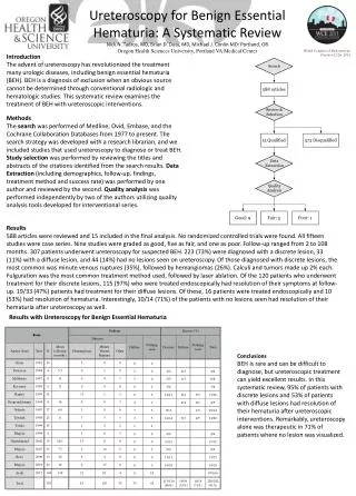
EVALUATION OF LATERALIZING ESSENTIAL HEMATURIA • Lateralizing essential hematuria, which has also been called chronic unilateral hematuria or benign essential hematuria, is an uncommon syndrome characterized by intermittent or continuous gross hematuria that cannot be diagnosed by radiologic and hematologic tests • Only cystoscopic examination can positively establish that blood is coming from one of the upper collecting systems
OTHER APPLICATIONS • Ureteroscopy should be performed after an upper tract localization study to detect concealed malignant lesions • Another major diagnostic application of ureteroscopy is surveillance following conservative treatment of upper urinary tract tumors. It is often difficult to differentiate radiographically between the recurrence of malignancy and normal postoperative scarring. Ureteroscopy with or without biopsy provides an excellent method of checking previous segmental resection sites
A significant indication for ureteroscopy is to follow up on a recent endourologic strategy for treating lowgrade and low-stage upper-tract transitional cell carcinoma. Concerning the surveillance after endoscopic surgery, urinalysis, urine cytology, and excretory or retrograde pyelography are not adequate, and ureteroscopic evaluation with upper trace biopsy is essential
THERAPEUTIC INDICATIONS CALCULI • The treatment of calculi, or the removal of stones, from various regions remains the most common application of ureteroscopy • Ureteroscopy is particularly effective in the management of distal ureteral stones, and rigid ureteroscopy provides a stone removal rate of close to 100%
Fuchs’ guideline—size limits of 1.5 cm for solitary stones and < 5 mm for multiple stones (no more than five in number)—is reasonable for successful ureteroscopy • Renal stones associated with intrarenalstenosis, such as stones above infundibular strictures and in calicealdiverticula or stones associated with ureteropelvic junction (UPJ) obstruction, are also indications for ureteroscopy
FOREIGN BODIES • A foreign body in the upper urinary tract is not common, but normally can be diagnosed by clinical history and radiography • Most foreign bodies reported recently are migrated or fragmented double-pigtail catheters or broken parts of accessory devices such as portions of stone baskets
UPPER-TRACT NEOPLASMS • Therapeutic ureteroscopy as an option for the following types of patients: (1) those who have a solitary kidney, bilateral upper-tract malignancy, or renal insufficiency and are not candidates for dialysis or a transplant, and (2) those who are poor risks for open surgery
BENIGN LESIONS • Benign lesions such as fibroepithelial polyps, inverted papilloma, and hemangioma are well suited to ureteroscopic resection or fulguration, although these lesions are rare • For renal hemangiomas, which are most often present on the papillary tips (see Figure 26-5) and are the most important cause of lateralizing essential hematuria, ureteroscopic fulguration is absolutely the first choice of treatment
STRICTURES • Ureteral strictures can be treated by dilation or endoscopic incision through a retrograde, antegrade, or combined approach using rigid and/or flexible ureteroscopes FISTULAS • Ureteroscopy is the treatment of choice for the resolution of fistulas such as a ureterovaginal fistula following hysterectomy
Ureteroscopy is successful in more than 95 out of 100 people RISKS • Complications are more likely when the stone is close to the kidney (upper third of the ureter) and include: • Injury to the ureter • Urinary tract infection • Bleeding • Abdominal pain
RIGID VERSUS FLEXIBLEURETEROSCOPES • Rigid and flexible ureteroscopes are used in a complementary fashion to access the entire upper urinary tract • The rigid ureteroscopesare ideal for treating pathology in the lower ureter, particularly below the level of the iliac vessels, as this portion of the ureter can easily be accessed with this instrument in most patients • The advantages of rigid ureteroscopes include ease of introduction into the orifice under direct vision, excellent image transmission, and larger flow and working channels
Flexible ureteroscopes, on the other hand, are better suited for the upper ureter, renal pelvis, and calices. Flexible ureteroscopes are more difficult to use in the lower ureter because of their tendency to buckle into the bladder (though this can be overcome up to a point by passing them over a working guidewire to stiffen the shaft temporarily during insertion, before this working wire is sacrificed)
The advantages of the flexible ureteroscopes are their ability to safely negotiate the angulations of the ureter, and they can access the entire upper collecting system in over 90% of patients with active and secondary passive deflection, and now with secondary active deflection. • Expertise with both rigid and flexible ureteroscopy allows the urologist to more safely diagnose and treat problems anywhere inside the upper urinary tract collecting system Smiths Textbook of Endourology, 2nd ed.
CYSTOSCOPY INDICATIONS • Frequent urinary tract infections • Blood in your urine (hematuria) • Loss of bladder control (incontinence) or overactive bladder • Unusual cells found in urine sample • Need for a bladder catheter • Painful urination, chronic pelvic pain, or interstitial cystitis • Urinary blockage such as prostate enlargement, stricture, or narrowing of the urinary tract • Stone in the urinary tract • Unusual growth, polyp, tumor, or cancer
A systematic approach is required when evaluating the urethra, prostate, bladder walls, dome and neck, and ureteral orifices (including location, number, shape, and character of efflux). • The bladder should be evaluated at different levels of filling. It is only after full distention of the bladder that characteristic glomerulations and ecchymoses are seen in interstitial cystitis. • Rectal examination with the endoscope in place is informative, especially in assessing prostate size and length of prostatic urethra. • Similarly concurrent vaginal examination in women can be useful in evaluation of cystoceles • Endoscopic inspection allows for identification of calculi, foreign bodies and mucous plugs, and also has the potential for intubation of ureterointestinal anastomoses. Smiths General Urology, 17th ed.
URETHROSCOPY • To identify and aid in treating urethral pathology, endoscopic inspection via a urethroscope with a 0° lens is helpful • Stricture disease can be identified or confirmed after radiographic studies. Strictures are characterized by circumferential narrowing • Urethral diverticulum can be identified with urethroscopy. A catheter can be placed through the neck of the diverticulum to help confirm its location during definitive open surgical repair. • Urethroscopy can be used to direct injection of dye into rare retained mullerian duct cysts, to identify and extract foreign bodies or rare calculi, and to access biopsy-suspicious lesions. • Urethroscopy allows endoscopic treatment of urethral condylomata Smiths General Urology, 17th ed.