Download
1 / 17
170 likes | 604 Vues
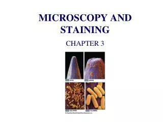
Microbiology Chapter 3 Microscopy and Staining. What’s on a Pinpoint?. How many bacteria? How many are needed to start an infection? Sometimes as few as 10 bacteria are enough!. Historical Microscopy. Anton van Leeuwenhoek-1670’s 1 st to see micro-organisms

E N D
What’s on a Pinpoint? • How many bacteria? • How many are needed to start an infection? • Sometimes as few as 10 bacteria are enough!
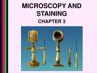
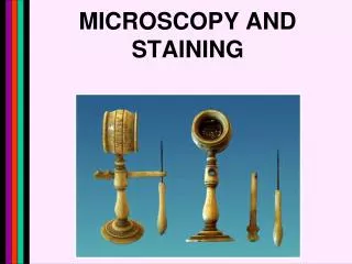
Historical Microscopy • Anton van Leeuwenhoek-1670’s • 1st to see micro-organisms • lens maker, simple scopes 100x to 300x • Single lens, like a magnifying glass • Studied “animalcules”
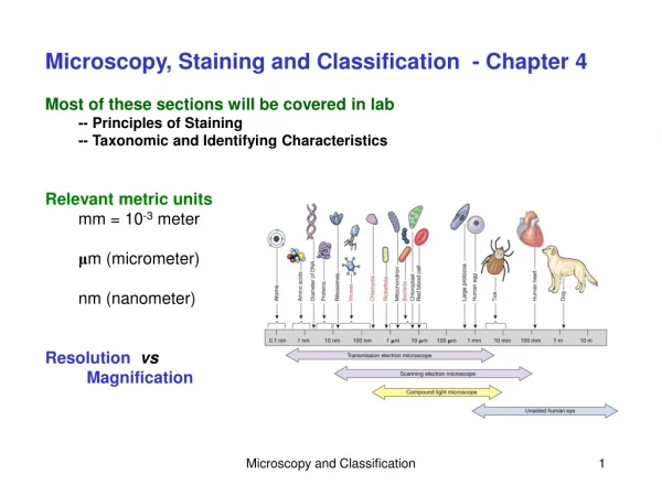
Principles of Microscopy • Metric units- powers of 10 • Microscopy- technology of making very small things visible to naked eye • Measurements in: - micrometers (microns) um 0.000001m= 10-6 m - nanometers nm= 10-9 m - angstroms- (A) 10-10 m
Properties of Light:Wavelength and Resolution • Wavelength- length of a light ray • Resolution- ability to see 2 objects as separate & discrete units (not fuzzy) • Visible light = 550nm (NG) • UV light= 100-400 nm better for resolution • Electron microscopy- .005 nm high reso • Resolving power of lens- numerical measure of lens, smaller distance from lens to slide =greater resolving power
Properties of Light:Light and Objects • Reflection-light strikes an object & bounces back • Transmission- light passes through object • Absorption- light rays taken up by object • Luminescence-absorbed UV rays are changed to longer wave & reemitted • Fluoresce- luminescence only occurring during irradiation • Phosphorescent- object emits light when light rays no longer strike it (some bacteria)
Properties of Light:Light and Objects • Refraction- bending of light as it passes from one medium to another • Index of refraction- measure of the speed at which light penetrates • Immersion oil- used for better resolution because oil as the same index of refraction as glass. • Diffraction- light waves bend around an opening and could cause blurry slides • Iimit = oil immersion with 10 x eyepiece=1000X
Light Microscopy and Types of Microscopes • Microscope that uses visible light to observe specimen • Hooke’s compound microscope had more than 1 lens • The Compound Light Microscope: - monocular- 1 eyepiece, binocular-2 • Survey of microscope parts and their functions – pg 58
Total Magnification Calculations • Scanning power -4x X 10x (ocular)= 40x • Low power 10x X 10x(ocular) = 100 x • High dry power 40x X 10 x = 400 X • Oil immersion 100x X 10 x = 1000x • Parfocal- in focus on one power, simple rotate nosepiece and its should focus on next power • Ocular micrometer- measure size of sample
Light Microscopy and Types of Microscopes • Dark-Field Microscopy- condenser causes light to reflect off specimen at an angle and increases the contrast • Phase-Contrast Microscopy-to observe live and unstained specimens by increasing refractive index and shows different degrees of brightness • Nomarski Microscopy- differential interference contrast and looks “3D”
Light Microscopy and Types of Microscopes • Flourescence- UV light is used to excite molecules, longer wavelengths= bright • Confocal Microscopy- usesbeams of UV lases light and computer reconstructs images, up to 40% better. Can study microbes alive or not. • Digital Microscopy-have built in digital camera and can be viewed on screen
Different Types of Electron Microscopy • EM uses electron beam and electro-magnets not lenses- high resolution • Photos taken – Electron micrographs • Transmission Electron Microscopy- (TEM) better view of internal structures up to 500,000x magnification - shadow casting- - freeze fracturing- - freeze etching-
Different Types of Electron Microscopy • Scanning Election Microscopy (SEM)- - Image of the surface “3D” 50,000x mag • Scanning Tunneling Microscopy (STM’s)- - 1980 can be used with liver specimens and under water • Atomic force microscope-(AFM)- advanced 3d from atomic size to 1 micron - used to study DNA, proteins
Techniques of Light Microscopy • Preparation of Specimens for the Light Microscope: • 1) Wet Mounts- drop of medium with microbes is spread on a slide • 2) Smears- microbes from a loopful of medium are spread on a slide, then heat fixed to kill microbes - heat fixation-
Principles of Staining • Stain- dye that binds to a cellular structure and gives it color • + charge-basic= methylene blue, crystal violet, safranin and malachite green • - charge-acidic= eosin and picric acid • Simple stain- single dye and reveals basic cell shapes and structures • Differential stain- 2 or more dyes: Gram stain, Ziehl-Neelsen acid fast and spore
Gram Stain • Gram Stain- 1884 crystal violet (+) and iodine and ethanol decolorizer, and counterstained with safranin (-) • Gram +=purple • Gram - = red • Gram non reactive= no stain • Gram Variable= stain unevenly
Special Staining Procedures • Ziehl-Neelsen Acid-Fast Stain - 1882 modification of Ehrlich staining method - Acid fast retain red color in cell walls • Negative staining-capsule is present and won’t take up stain • Flagellar staining- coats flagella so they can be seen • Endospore staining- Schaeffer-Fulton stain