Download
1 / 43
430 likes | 443 Vues
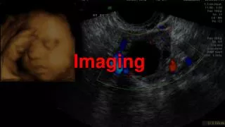
Alternative Imaging. Chapter 21. Introduction. Other modalities have assumed prominence in imaging field, especially for diagnosis of diseases that are often difficult to see on radiographs. Imaging techniques are complementary to radiography. Ultrasonography.

E N D
Alternative Imaging Chapter 21
Introduction • Other modalities have assumed prominence in imaging field, especially for diagnosis of diseases that are often difficult to see on radiographs. • Imaging techniques are complementary to radiography.
Ultrasonography • Seen in veterinary medicine since the 1980’s. • Can provide information about organ architecture independent of organ function. • Helpful in debilitated or young patients in which contrast agents or exploratory surgery is contraindicated.
Technical Aspects • Ultrasound beam is created by a piezoelectric crystal that oscillates at several million Hertz per second (mHz) within a tranducer (probe). • When sound wave interacts with tissues of the body, it is reflected and echo is received by the tranducer. • Ultrasound machines display images in real time.
Terminology • Acoustic impedance- Relationship between density or stiffness of tissue and the velocity of sound within the tissue. Differences in acoustic impedance of adjacent tissues determine the intensity of the reflected sound. • Attenuation- Reduced intensity of radiation caused by absorption or scattering, or both, during passage through tissue. Sound is also attenuated as it passes through tissue and the intensity is reduced.
More Terminology… • Distant enhancement- Ultrasound artifact. Increased sound intensity beyond a fluid-filled, anechoic area, created by absence of attenuation of the sound beam as it passes through the fluid. • Acoustic Shadowing- Ultrasound artifact. Echo-free zone created distal to the imaged organ when sound waves hit a highly reflective tissue that prevents sound from being transmitted to greater depths.
And more terminology…. • Echogenicity-Intensity of reflected echoes. • Anechoic- No echoes are detected, and the area is black. Typically associated with fluid-filled structures such as the urinary bladder. • Hypoechoic- A few echoes are detected, and the area is low-level gray compared with adjacent tissues. Usually seen with solid homogenous tissues or complex fluid containing cells such as blood.
And one more…. • Hyperechoic- Echoes produced are brighter than in surrounding tissue.
Clinical Applications • Echocardiography • Used to evaluate cardiac disease. • Two modes: • M-mode (motion mode)- information is displayed as depth versus time on graph. • B-mode (brightness mode)- Intensity of returning echoes is expressed as brightness in the display. • No specific preparation, chest wall is clipped, gel applied and animal is restrained. • Must work between ribs of chest wall. • Difficult in deep chested animals.
Echocardiography continued. • Long axis view- Echocardiographic image showing the heart from base to apex in a longitudinal or sagittal plane. • Short axis view- Echocardiographic image showing the heart in transverse plane. • Doppler shift-Difference between transmitted and received sound frequencies. The greater the Doppler shift, the greater the flow velocity.
Abdominal Ultrasound • Patient must be fasted for 12 hours to reduce the amount of intestinal gas. • Full urinary bladder is optimal for scanning the bladder or prostate. • Liver and biliary tract: • Radiographs are superior to ultrasound for assessing liver volume. • Ultrasound-guided biopsy or fine-needle aspiration is often performed in conjunction with liver scanning. (Sedation of anesthesia is required).
Spleen • Normal spleen is elliptic, flat, and smoothly contoured. • Pancreas • Normal pancreas is narrow, smoothly marginated and hypoechoic. • Pancreatitis is most common indication for scanning.
GI tract • Can be difficult due to variable amounts of gas within the lumen, which reflect sound and prevent imaging of deeper structurs. • Can be used to identify masses, assess bowel peristalsis, foreign bodies, and confirm intussusception.
Kidneys • Normal kidney has hyperechogenic capsule. • Ultrasonography is used when kidneys may not be visualized on radiographs, or to assess location and distribution of disease in enlarged kidneys. • Does not assess kidney function unless doppler technique is applied. • Helpful to identify fluid-filled, cystlike lesions or solid masses. • Biopsy usually needed to confirm diagnosis.
Adrenal glands • Normal adrenal glands are small and located in the perirenal fat medial to the cranial pole of each kidney. • Left adrenal gland has dumbbell shape while right adrenal gland is more triangular. • In hyperadrenocorticism, both glands become enlarged but there is no change in shape. • In cases of masses, usually on one side and alters the shape of the gland.
Prostate • Normal prostate has homogeneous echogenicity and fine texture. • Indicated for cases of prostatomegaly, signs of lower urinary tract disease, constiplation, and caudal abdominal pain. • Can not differentiate between benign hyperplasia and neoplasia so biopsy would be recommended.
Urinary Bladder • Normal bladder contains anechogenic urine. • Bladder is smooth and wall is uniform in thickness. • Indicated with potential lower urinary tract disease, stones, masses, etc. • Urethral masses are not visible because are hidden by pelvis.
Reproductive Tract • Normal reproductive tract in nonpregnant animals is not normally seen. • Indications are to diagnose pregnancy, pyometra, stump granuloma, or ovarian neoplasia. • Pregnancy detection is 30 days after breeding.
Eyes • Can place transducer on cornea. • Can visualize components of the eye and scan for intraocular masses or hemorrhage. • Extremities • Focused primarily on equine limb below the carpus and tarsus.
Computed Tomography (CT) • One of the most expensive diagnostic tests in veterinary medicine. • Advantage is can acquire information not available from radiographs, contrast studies, or ultrasound examination. • Indications are for central and peripheral nervous system diseases of the brain, spinal cord, and lumbosacral spine. • Also useful for obscured masses in the mediastinum, axillary region, and retroperitoneal space.
Technical Aspects • CT uses x-rays and computers to produce images that show anatomy in cross section. • Allows for visualization of structures in sagittal, dorsal, transverse, and oblique planes without superimposition artifact from fat, ribs, spine, pelvis, or any organs that may mask detail on a survey radiograph. • Consists of a movable bed or cradle on which patient lies and a gantry that contains the x-ray tube and detectors. • Tube and detectors can be moved 360 degrees. • Generally requires general anesthesia.
CT Terminology • Pixels (picture elements)- Tiny squares making up the image matrix; represent voxels. • CT number- Number converted to gray scale in the final image, which represents the attenuation of the x-ray beam in tissue within a voxel. The number is also referred to as a Hounsfield number, named for the inventor of CT scanning.
Clinical Applications • Skull • Radiographs are generally not extremely diagnostic of this region. • Lesions can be easily seen by CT. • Indications for skull CT are seizure, blindness, vestibular signs, and change in disposition.
Spine • CT is helpful with myelography to define lesions on spine. • Method of choice for imaging spine caudal to L4-5. • Can allow visualization of intervertebral disk potrusion.
Extremities • Assess coronoid processes in dogs. • Scan both elbows because may be bilateral. • May also be used to visual meniscal disease, osteochondrosis, and sequestra. • Thorax • Indicated with pulmonary and mediastinal masses, and can evaluate metastasis.
Abdomen • Liver, gallbladder, stomach, small intestines, pancreas, spleen, adrenal glands, kidneys, ureters, urinary bladder, prostate, ovary, colon, and major vessels are easily identified on CT scans. • Useful for canine adrenal masses.
Nuclear Scintigraphy • Noninvasive imaging procedure that uses a small amount of radioactive material administered intravenously, transcolonically, or by aerosol insufflation. • Do provide physiologic information about the function of specific organs.
Technical Aspects • Radioactive isotope that emits predominantly gamma rays. • Gamma camera detects the gamma emissions from the radionuclide and forms a black and white image of the selected organ printed on x-ray film.
Terminology • Labeled Compounds- A compound whose molecule is tagged with a radionuclide. • Radiopharmaceutical- A radioactive drug that can be administered for diagnostic or therapeutic purposes. • Half-life (t1/2)- Time in which the initial activity of a radionuclide is reduced to one half. Biologic half-life includes exertion, as well as the characteristic half-life of the isotope. • Target organ- The organ intended to be imaged and expected to receive the greatest concentration of administered radioactivity.
Clinical Applications • Thyroid • Evaluates for hyperthyroidism. • Image shows a blackened area in the involved lobe of the thyroid gland. • Bone • Indicated for lameness that cannot be localized by physical exam. • Liver • Indicated in patients with a small liver or evidence of a liver mass, decreased liver function, biliary outflow obstruction, or abnormal hepatic blood flow. • Looking for portosystemic shunt.