Download
1 / 24
240 likes | 403 Vues
Lecture 5: Cellular Level Methods. So far we’ve seen some methods for assessing the chemical and/or physical state of a protein.

E N D
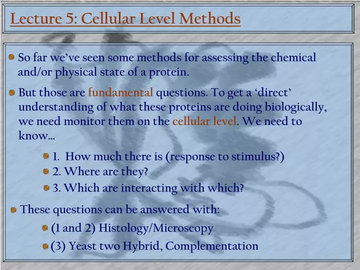
Lecture 5: Cellular Level Methods So far we’ve seen some methods for assessing the chemical and/or physical state of a protein. But those are fundamental questions. To get a ‘direct’ understanding of what these proteins are doing biologically, we need monitor them on the cellular level. We need to know… 1. How much there is (response to stimulus?) 2. Where are they? 3. Which are interacting with which? These questions can be answered with: (1 and 2) Histology/Microscopy (3) Yeast two Hybrid, Complementation
How much is there? Remember, most of the methods we’ve talked about for observing proteins are not quantitative. So no matter what happens, we are likely only going to be able to get a relative answer. Effectively, this will limit us to answering the ‘how much’ question for stimulated or ‘disease state’ versus ‘normal state’ cells. But before we can do any of that, we need to: Get the label in and attached to the correct protein Get the cells in a state where they can be observed
Getting the Label in: Histology Very often, the molecular biology required to transform eukaryotic cells is prohibitive. An alternative option is ‘fix’ the cell at a certain time and then label it. This is cellular level histology. 1. Grow cells under desired conditions 2. ‘Fix’ cells in a tissue sample Done with ‘formalin’ (formaldahyde, water, methanol). Crosslinks proteins by forming methylene bridges Embed cells in parafin wax. 3. Cut thin ‘slices’ of wax embedded tissue. Dry on to cover slip
Histology: Chemical Stains You are now ready to attach your probe The older probes are dyes that bind to specific regions or organelles for visualization: H&E (Hematoxylin/Eosin) Stain Wright Stain for Immune Cells
Histology: Immunohistochemistry Chemical Stains are non-specific. They rarely target a specific protein. Immunohistochemistry uses modified antibodies to target specific proteins/molecules. Polyclonal antibodies are made by injecting an animal with your target analyte. Monoclonal antibodies are made by injecting an animal with your target analyte. spleen cells Myeloma cells HGPRT- Immune response fused
Immunohistochemistry So these immunoglobulins will ‘stick’ to the antigens against which they are raised in our fixed cells. But how do we see them? Of course we’re going to add a chromophore. But why modify every antibody you make when you can create a generic ‘secondary antibody’ directed at the unchanging part of the ‘primary’ antibody: We can direct antibodies at the ‘constant’ part of the heavy chain
Biotin/Strepavidin/HRP Detection One of the first, and currently most commonly used detection systems is… Streptavidin Biotinylated Horseradish Peroxidase 2° Antibody H2O2
Immunohistochemistry Examples So histology and immunohistochemistry can tell us which cells… Are producing how much protein Prion Protein PAX5 HBcAg Beta III tubulin (neurone specific) CD3, CD20 NIH 3T3
In Situ Hybridization But, what if we can’t make an antibody or the target protein is inaccessible? cRNA w/ probe Target Protein mRNA
In Situ Hybridization Examples In Situ Hybridisation is a little more specific, allowing us to quantitate within cells, but mostly still used at tissue level CD5 Chromosome 1
Histology Instrumentation For Processing For Staining For Cutting (microtome)
Getting the Label In: Chimeras To make a Chimera, the gene encoding the protein of interest is modified to encode the analyte plus the reporter Restriction Enzyme site Stop Target protein Promoter P Target Linker (poly-G) P EGFP Target Same promoter = same level of production!?
In Cell Localization: Fluorescence Fluorescence/Immunohistochemistry is the most commonly used tool to localize proteins at the sub-cellular level. Actin endoG-YFP (apoptotic endonuclease) Mitochondria Debrin Colocalization Apoptosis 12 (7): 1155-1171, 2007
Instrumentation: Confocal Microscopy In confocal microscopy, the illuminating light is focused on a tiny section of the sample. The primary advantage of confocal microscopy is that it eliminate any light that is not from the focal plane of the focusing lens (which would be out of focus).
Outside the Cell: The Western Blot ‘Western blots’ are basically Immunohistochemistry outside the cell Bust it open! Nitrocellulose Membrane 2° (skim) Electrophoresis All extract proteins on membrane 1°
Outside the cell: Antibody Microarrays If you want to analyze the proteome in parallel… This method is semi-quantitative. You can use a known concentration of antigen as a standard.
What Sticks to What: The ‘Interactome’ One of the most pressing questions in biochemistry is protein function. You can tell a lot about what a protein does by figuring out what it interacts with. This – and not the gene level – is where the complexity of life arises: (admittedly, we humans do more with our genes than the roundworm via RNA splicing) Human genome?: 20,000-25,000 genes Roundworm Genome?: ~ 20,000 genes
Uncovering Protein/Protein Interactions One of the first methods for uncovering Protein/Protein interactions was the ‘yeast-two-hybrid’ screen Any method used must be parallel Analyte proteins are overexpressed with Gal4 AD and BD UAS Promoter binders Must be able to get into the yeast nucleus Weak, transient interactions can still activate reporter Consequently, Y2H screens are considered low confidence
Phage Display Phage Display relies on the ‘display’ of a peptide sequence on the C-terminus of a phage coat protein (pIII, IV or 10B) These are made to interact with a ‘library’ of immobilized proteins or peptides Can use unnatural selection to amplify good binders
Phage Display and Yeast 2 Hybrid Both Phage Display and Yeast Two Hybrid can produce extremely complicated interaction maps, if the genome is well known
Phage Display and Yeast 2 Hybrid But both these techniques have high rates of false positives, so… Science 295 No.5553(2002): p321-4 Phage Display PD = 369 Interactions Y2H = 233 Interactions 59 Interactions Y2H
Protein Microarrays In protein microarrays, proteins are ‘printed’ (literally) onto a glass slide… This microarray has every protein in the S. Cerevisiae genome Proteomics (2003); 3(11):2190-9. A ‘liver protein’ microarray Proteomics 7 (13): 2151-2161 2007 Proteins are detected in ‘duplicate spots’ to limit false positives
Protein Complementation Protein complementation is the least versatile protein interaction detection technique, but it may be the coolest… Proteomics 7 (7): 1023-1036, 2007 Nat. Meth. 4 (5): 421-427, 2007
Time-Resolved Localization Fluorescent labels can be used in living cells to monitor protein localization in real time. Apoptosis 12 (7): 1155-1171, 2007 BBRC 364 (2): 231-237, 2007