Download
1 / 38
380 likes | 584 Vues
CATARACT IN ADULT EYE. Prepared by : DR BADR AL AHMADI. Supervised by : DR KHALID AL ARFAJ. Introduction. DEFINITION : cataract is a degradation of the optical quality of the crystalline lens PATIENT POPULATION DEFINITION Adults (18 years old and older) with cataracts.

E N D
CATARACT IN ADULT EYE Prepared by : DR BADR AL AHMADI Supervised by : DR KHALID AL ARFAJ
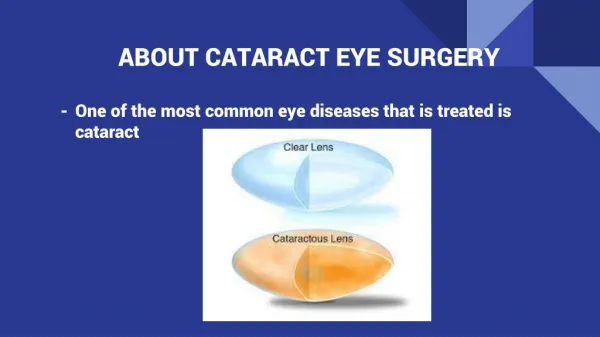
Introduction • DEFINITION : cataract is a degradation of the optical quality of the crystalline lens • PATIENT POPULATION DEFINITIONAdults (18 years old and older) with cataracts.
CLINICAL OBJECTIVES • Identify the presence and characteristics of a cataract • Assess the impact of the cataract on the patient’s visual and functional status and on quality of life • Educate the patient about the impact of a cataract on vision, functional activity, and natural history as well as the benefits and risks of surgical and other alternatives so that the patient can make an informed decision about treatment options • Establish criteria for a successful treatment outcome with the patient • Perform cataract surgery when there is the expectation that it will benefit the patient’s function and when the patient elects this option • Perform surgery when indicated for management of coexistent ocular disease • Provide necessary postoperative care, rehabilitation, and treatment of any complications
PREVALENCE • Cataracts are the leading cause of treatable blindness worldwide and remain an important cause of blindness and visual impairment in the United States, accounting for approximately 50% of visual impairment in adults over the age of 40. • Cataract affects over 22 million Americans age 40 and older, or about 1 in every 6 people in this age range. By age 80, more than half of all Americans have cataracts. • The Eye Diseases Prevalence Research Group estimated that the number of individuals with cataracts will increase by 50% by 2020.
PREVALENCE Types of cataracts : • Nuclear : consist of a central slowly progressing opacification that affect distance vision more than near vision. More common in an older population • Cortical : can be central or peripheral and sometimes are best visualized by retroillumination or retinoscopy. Patients with this type of cataract commonly complain of glare. When the entire cortex becomes white and opaque, the cataract is referred to as a mature cortical cataract.
PREVALENCE Types of cataracts : • Posterior subcapsular (PSC) cataracts can cause visual impairment if they affect the axial region of the lens. Posterior subcapsular cataracts are found more often in younger patients than are nuclear or cortical cataracts. Patients often have glare and poor vision with bright lighting, and their near vision is typically more affected than distance vision
PREVALENCE • In the Salisbury Eye Evaluation Study, Americans of African descent had a four times greater chance of having cortical opacities than Americans of European descent, and Americans of European descent were more likely to have nuclear and PSC opacities. • The Los Angeles Latino Eye Study of individuals 40 years old or older found that cortical opacities were the most frequent type of lens opacity
NATURAL HISTORY • With age, the lens increases in thickness and weight. • Cataract is a progressive disease. Once visual acuity and function start declining, the natural history is a steady decline without any chance of recovery. • In the Barbados Eye Studies, individuals with pre-existing lens opacities had cumulative 9-year progression rates of 22.0% for cortical, 17.8% for nuclear, and 25.8% for PSC opacities. • The Melbourne Visual Impairment Project reported cumulative 5-year progression rates of 14.3% for cortical, 19.3% for nuclear, and 20.0% for PSC opacities.
PREVENTION • Several studies show a linkage of smoking with nuclear sclerosis and demonstrated a benefit from smoking cessation. • Cumulative lifetime exposure to ultraviolet-B radiation has been associated with lens opacities; therefore, brimmed hats and ultraviolet-B blocking sunglasses are reasonable precautions to recommend to patients. • A systematic review and eight randomized controlled trials of nutritional or vitamin supplementation showed no significant effect in delaying the onset or progression of cataracts.
PREVENTION • Patients with DM are at higher risk for cataract formation, and so prevention and proper treatment of type 2 diabetes may have the additional benefit of reducing the risk of cataract. • In general, prevention is to get away of risk factors.
VISUAL FUNCTION AND QUALITY OF LIFE Improved visual function as a result of cataract surgery can be characterized by the following: • Better optically corrected vision • Better uncorrected vision with reduced eyeglass dependence • Increased ability to read or do near work • Reduced glare • Improved ability to function in dim levels of light • Improved depth perception and binocular vision by elimination of anisometropia and achievement of good functional acuity in both eyes • Improved color vision
VISUAL FUNCTION AND QUALITY OF LIFE Improved physical function as a critical outcome of cataract surgery can be characterized by the following: • Increased ability to perform activities of daily living • Increased ability to continue or resume an occupation • Increased mobility (walking, driving)
VISUAL FUNCTION AND QUALITY OF LIFE Improved mental health and emotional well-being as a second critical outcome of cataract surgery includes the following benefits: • Improved self-esteem and independence • Increased ability to avoid injury • Increased social contact and ability to participate in social activities • Relief from fear of blindness
Care Process PATIENT OUTCOME CRITERIA • Reduction of visual symptoms • Improvement in visual function • Achievement of desired refractive outcome • Improvement in physical function, mental health, and quality of life
Evaluation of Visual Impairment • There is no single test or measure that adequately describes the effect of a cataract on a patient's visual status or functional ability. Similarly, no single test can properly define the threshold for performing cataract surgery. • The Snellen visual acuity chart is an excellent method for testing distance refractive error
Evaluation of Visual Impairment • Testing distance vision with high-contrast letters viewed in dark-room conditions will underestimate the functional problems in common real-life situations. • Because preoperative visual acuity alone may be an unreliable predictor of postoperative functional improvement, the decision to recommend cataract surgery should not be made solely on the basis of Snellen visual acuity.
Ophthalmic Evaluation • Patient history, including the patient's assessment of functional status, medical conditions, medications currently used, and other risk factors that can affect the surgical plan or outcome of surgery (e.g., immunosuppressive conditions, systemic alpha-1 antagonists, diabetes) • Visual acuity with current correction at distance and, when appropriate, at near • Measurement of best-corrected visual acuity (with refraction when indicated) • External examination (eyelids, lashes, lacrimal apparatus, orbit)
Ophthalmic Evaluation • Examination of ocular alignment and motility • Assessment of pupillary function • Measurement of intraocular pressure (IOP) • Slit-lamp biomicroscopy of the anterior segment • Dilated examination of the lens, macula, peripheral retina, optic nerve, and vitreous • Assessment of relevant aspects of the patient's mental and physical status
Supplemental Ophthalmic Testing • Ophthalmologist is able to determine whether the cataract is responsible for the patient's visual loss by correlating slit-lamp biomicroscopy findings with the patient's specific symptoms. • Visual acuity testing alone does not quantify certain visual symptoms, such as disabilities due to glare and reduced contrast sensitivity • Glare testing determines the degree of visual impairment in the presence of a light source located in the patient's visual field.
Supplemental Ophthalmic Testing • Visual acuity in some patients with cataracts may be normal or near normal when tested in a dark examination room, but when these patients are retested together with a source of glare, visual acuity (or contrast sensitivity) may drop significantly. • Contrast sensitivity testing measures the eye's ability to detect subtle variations in shading by using figures that vary in contrast, luminance, and spatial frequency. • Ocular wavefront testing has demonstrated that even relatively mild cataracts may be associated with a significant increase in visual aberrations.
Supplemental Ophthalmic Testing • Potential acuity testing attempts to predict the visual acuity following cataract surgery. • Potential Acuity Meter, laser interferometer, and scanning laser ophthalmoscope project an image onto the retina through relatively clear regions of the lens, and the patient is asked to identify the letters or pattern.
Supplemental Ophthalmic Testing • Electrophysiologic testing (e.g., ERG & VEP) measures the electrical response to visual stimuli presented and indicates potential retinal function in nonverbal patients. • Specular microscopy and corneal pachymetry have been used to evaluate patients with known preoperative corneal disease in an effort to determine whether the cornea is likely to remain clear following cataract surgery.
Supplemental Ophthalmic Testing • Optical coherence tomography (OCT) and fluorescein angiography may be helpful prior to cataract surgery for confirming normal foveal architecture or for identifying the presence of comorbid disease. • B-scan ultrasonography is appropriate when a dense cataract precludes adequate visualization of the posterior segment or to confirm the presence of a posterior staphyloma. • Visual fields, external and fundus photography, and special color-vision testing have not been shown to be of value in routinely evaluating patients before cataract surgery.
Nonsurgical Management • Counseling patients about cataract-related visual symptoms, providing reassurance about the cause of the visual disability, and prescribing new eyeglasses where appropriate. • Reassurance of the patient by reducing their risk of cataract development or progression by modifying their exposure to risk factors.
Indications for Surgery • The primary indication for surgery is visual function that no longer meets the patient's needs. • Clinically significant anisometropia in the presence of a cataract • The lens opacity interferes with optimal diagnosis or management of posterior segment conditions • The lens causes inflammation or secondary glaucoma (phacolysis, phacoanaphylaxis) • The lens induces angle closure (phacomorphic)
Contraindications to Surgery • Tolerable refractive correction provides vision that meets the patient's needs and desires • Surgery is not expected to improve visual function, and no other indication for lens removal exists • The patient cannot safely undergo surgery because of coexisting medical or ocular conditions • Appropriate postoperative care cannot be arranged • The patient or patient's surrogate decision maker is unable to give informed consent for nonemergent surgery
Preoperative Medical Evaluation • To examine the patient preoperatively . • To ensure that the documented evaluation accurately reflects the symptoms, findings, and indications for treatment • To obtain informed consent from the patient or the patient's surrogate decision maker after discussing the risks, benefits, and expected outcomes of surgery, including the anticipated refractive outcome and the surgical experience. • To review the results of the presurgical evaluation with the patient or the patient's surrogate decision maker • To formulate a surgical plan, including selection of an appropriate IOL • To formulate postoperative care plans and inform the patient or the patient's surrogate decision maker of these arrangements (setting of care, individuals who will provide care) • To answer the patient's questions about the surgery and care, including associated costs
Preoperative Medical Evaluation • All patients undergoing cataract surgery should have a history and physical examination relevant to the risk factors for undergoing the planned anesthesia and sedation and as directed by a review of systems. • patients with certain systemic diseases, a preoperative medical evaluation by the patient's primary care physician should be strongly considered.
Biometry and IOL Power Calculation • The accurate measurement of axial length and central corneal power, combined with an appropriate IOL selection based on a power calculation formula, is the minimal requirement to achieve the targeted postoperative refraction. • A-scan ultrasonography or optical biometry is used to measure axial length
Biometry and IOL Power Calculation • Formulas for calculating IOL power rely on keratometry to determine the net refractive contribution of the cornea. These measurements can be obtained by either manual or automated keratometry, or through corneal topography. • The surgeon should consider the patient's individual desires and needs in selecting an appropriate postoperative refractive target.
optical biometry A-scan ultrasonography • Non contact method. • Light source. • Operator independent. • More accurate. • Measure refractive A.L. • Better when silicon oil in posterior segment. • Difficult in dense cataract or when Pt is unable to fixate properly. • Measure center of the macula when proper fixation is achieved. • contact method. • Ultrasonic source. • Operator dependent. • Less accurate. • Measure anatomical A.L. • Difficult when silicon oil in posterior segment. • Better in dense cataract or when Pt is unable to fixate properly. • Cannot Measure center of the macula
Anesthesia • general and local (regional) anesthesia (e.g., retrobulbar, peribulbar, sub-Tenons injection, topical, and intracameral). • The planned mode of anesthesia should be discussed with the patient so that she or he will know what to expect in terms of pain, discomfort, consciousness level, visual experiences, and complications. • Local (regional) anesthesia is generally preferred, with or without sedation/analgesia.
Anesthesia • Anesthesia techniques with needle injections may be associated with complications such as strabismus, globe perforation, retrobulbar hemorrhage, intravascular or subarachnoid injection, and macular infarction. • The risk of globe perforation is increased with axial myopia and following scleral buckle placement. • Monitoring during administration of anesthesia and surgery generally includes ECG, pulse oximetry, BP, and respirations. • In summary, the type of anesthesia management should be determined according to the patient's needs and the preference of the patient, the anesthesia professionals, and the surgeon.