Download
1 / 80
820 likes | 955 Vues
Organization of the CNS I. Phrenology. Review of the Nervous System. MAJOR FUNCTION: COMMUNICATION. Neuronal Morphology. The Neuronal Synapse. Glia – the Particular Role of Astrocytes. Astrocyte. Astrocytes Help Form the Blood Brain Barrier.

E N D
Review of the Nervous System MAJOR FUNCTION: COMMUNICATION
Astrocytes Help Form the Blood Brain Barrier • Basal lamina of the astrocytes + the astrocytic endfeet produce help maintain the BBB. • Notice how astrocytes send processes to the external surface of the CNS where the endfeet form the glia limitans externa, which separate the pia mater from the nervous tissue. • Gap junctions and desmosomes join the endfeet to form a space between neurons and vascular endothelial cells (Fig. 2.2).
General Orientation Terms • Rostral • Caudal • Posterior • Anterior • Dorsal • Ventral • Sagital • Horizontal • Coronal
Terms of Orientation:
Planes of Section: Horizontal Transverse (Coronal) Saggital
Major regions and landmarks • Six regions in the adult brain • Cerebrum • Diencephalon • Mesencephalon • Pons • Cerebellum • Medulla oblongata • Brain contains extensive areas of neural cortex • Layer of gray matter on the surface of the cerebellum and cerebrum
A. Spinal Cord • Conduit for flow of information from PNS to CNS. • Participates directly in body movement control. • Processes sensory information from limbs, trunk, and internal organs. • Has segmental organization (most like primitive or early developmental NS). • Each segment has a pair of nerve roots.
B. Brainstem and Cerebellum • Brainstem = medulla + pons + midbrain. • ‘spinal cord’ for the head (sensory, motor control). -use of cranial nerves in place of spinal nerves. b. Transmission of information between brain and SC. • Regulation of arousal (via reticular formation at the core of brainstem). • Regulation of important visceral functions (e.g., bp and respiration).
Brain Stem: Ventral Surface
Brain Stem: Dorsal Surface
B. Brainstem and Cerebellum • Cerebellum – regulation of movement, maintenance of posture and balance. -Works closely in concert with pons (derived from same embryonic brain division).
The Cerebellum • Adjusts postural muscles and tunes on-going movements • Cerebellar hemispheres • Anterior and posterior lobes • Vermis • Flocculonodular lobe • Superior, middle and inferior cerebellar peduncles link cerebellum with brain stem, diencephalon, cerebrum, and spinal cord • Interconnects the two cerebellar hemispheres
C. Diencelphalon • Diencephalon – 2nd only to cerebrum as the most highly developed division. • Thalamus – key for transmitting information to cerebral hemispheres. • Hypothalamus – integrates functions of the ANS – controls endocrine hormone release from pit.
The Thalamus • Final relay point for ascending sensory information • Coordinates the activities of the cerebral cortex and basal nuclei
Thalamus • Part of diencephalon – relay nuclei through which sensory and motor information pass to and from the cortex. • E.g., in the 2 1° systems we have discussed: - from sc 1° sensory cortex - from 1° motor cortex sc. • This is also the case for neural signals controlling other functions, such as learning and memory and emotions – and projecting to other parts of the cortex. A. Relay Nuclei have distinct roles, transmitting info from particular subcortical inputs to a specific portion of the cerebral cortex.
D. Cerebral Hemispheres • These structures mediate the most complex and sophisticated human behaviors. • Cerebral cortex – highly convoluted to accommodate large surface area: gyri, sulci, fissures – deeper grooves – often separate major divisions. 4 lobes:
Frontal • Motor behavior • The 1° cortex at precentral gyrus. -nearby premotor areas -prefrontal asociation cortex -cingulate gyrus - important for reasoning and emotional control.
Parietal • Sensory areas. • The 1° cortex at postcentral gyrus. -Superior parietal lobule -spatial perception, self-image -Inferior parietal lobule -integrating sensory information for speech and perception
Occipital • The 1° cortex within calcarine fissure on medial surface. • Surrounding association cortex elaborates the sensory message so that we can see and integrate forms and colours. • Situated at the division of parietal and occipital lobes is an area important for recognizing faces.
Temporal • Sensory functions, plus memory and emotions. • The 1° auditory cortex: on superior temporal gyrus – speech centre. • Much integration with nearby areas: -inferior temporal gyrus – perception of visual forms and colours. - works with nearby occipital. -temporal pole + medial temporal areas – mediate emotions along with nearby frontal cortical areas.
Ventricles of the Brain • Central passageway of the brain enlarges to form ventricles • Contain cerebrospinal fluid (CSF)
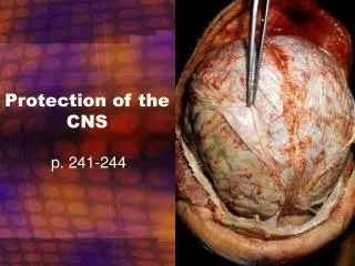
The Cranial Meninges • Continuous with the three layers of the spinal cord • Folds of dura mater help stabilize the position of the brain • Falx cerebri • Tentorium cerebelli • Falx cerebelli
Meninges of the CNS • 3 layers: • Dura - thickest • Arachnoid – “spider-like” • Pia – delicate, adheres to surface. • Protective • Circulating function (contain blood vessels) – many veins and arteries in subarachnoid space. Dural sinuses are major venous areas carrying blood away from the brain.
The Mesencephalon • The tectum (roof) contains the corpora quadrigemina • Superior and inferior colliculi • The mesencephalon contains many nuclei • Red nucleus • Substantia nigra • Cerebral peduncles • RAS headquarters
The Diencephalon is Composed of • Epithalamus • Hypothalamus • Thalamus