Download
1 / 26
270 likes | 347 Vues
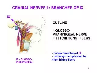
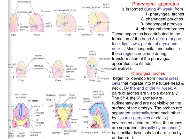
Pharyngeal apparatus It is formed during 4 th week from 1- pharyngeal arches 2- pharyngeal pouches 3- pharyngeal grooves 4- pharyngeal membranes

E N D
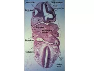
Pharyngeal apparatus It is formed during 4th week from 1- pharyngeal arches 2- pharyngeal pouches 3- pharyngeal grooves 4- pharyngeal membranes These apparatus is contributed to the formation of the head & neck ( tongue, face, lips, jaws, palate, pharynx and neck . . Most congenital anomalies in these regions originate during transformation of the pharyngeal apparatus into its adult derivatives Pharyngeal arches begin to develop from neural crest cells that migrate into the future head & neck . By the end of the 4th week, 4 pairs of arches are visible externally. The 5th & the 6th arches are rudimentary and are not visible on the surface of the embryo. The arches are separated externally, from each other by fissures ( grooves or clefts ) covered by ectoderm. Also, the arches are separated internallyby pouches ( balloonlike diverticula that are lined by endoderm ).
The first pair of arches ( the primordium of the jaws) appears as surface elevations lateral to the developing pharynex. The other arches appear as rounded ridges on each side of the future head & neck . . The 1st arch is called mandibular arch , from it two prominences are developed The maxillary prominence gives rise to the maxilla( upper jaw), Zygomatic bone ; Squamous part of the temporal bone . The mandibular prominence forms the mandible ( lower jaw The 1st pair plays a major role in facial development. These arches support the lateral walls of the primordial pharynx, which is derived from the cranial part of the foregut .
A 20 – week fetus illustrating the area of the face derived from the first pair of the pharyngeal arches. Derivatives of maxillary prominence Maxilla Palatine bone Zygomatic bone Squamous temporal bone
The primordial mouth or Stomodeum appears as a slight depression of the surface ectoderm . It is separated from the cavity of the primordial pharynx by a bilaminar membrane (oropharyngeal membrane ) It is composed of ectoderm externally & endoderm internally. It ruptures at about 26 days , thus the primordial pharynx& foregut become communicating with the amniotic cavity.
Pharyngeal arch Components Each arch consists of a core of mesenchyme and is covered externally by ectoderm & internally by endoderm. The original mesenchyme is derived from mesoderm in the third week. During the 4th week most of the mesenchyme is derived from neural crest cells which are the major source of the arches connective tissue ( bone, cartilage and ligaments) . A typical pharyngeal arch contains An aortic arch , an artery that arises from the truncus arteriosus of the primordial heart. ( vascular endothelial from the original mesenchyme ) . A cartilaginous rod that forms the skeleton of the arch . ( from the neural crest ) . A muscular component that differentiates into muscles of head & neck ( original mesen.) A nerve that supplies the mucosa & muscles. It is derived from neuroectoderm of the . Brain .
An aortic arch that passes around the primordial pharynx to enter the dorsa aorta .
Ventral parts of the 1st arch cartilage( mandibular process) form the horseshoe- shaped primordium of the mandible. The cartilage disappears as the mandible develops around it by intramembranous ossification of mesenchymal tissue surrounding it . The middle part regresses, but its perichondrium forms the ligaments. The dorsal end is closely related to the developing ear , it ossifies and form two middle ear bones malleus& incus ). The ventral end of the 2nd arch cartilage ossifies to form the lesser cornu and the superior part of the body of the hyoid bone The middle part regresses & its perichondrium forms styloid ligament. The dorsal end, ossifies to form stapes& styloid process. If stylohyoid ligament ossifies, it may cause pain in palatine tonsil . The ventral part of the 3rd arch cartilage, ossifies to form the greater cornu and the inferior part of the body of the hyoid bone. The 4th & the 6th arch cartilages fuse to form the laryngeal cartilages ,except the epiglottis. The 5th arch is rudimentary and has no derivatives.
The 1st arch forms muscles of mastication, tensor veli palatini, tensor veli tympani, mylohyoid and anterior belly of digastric. The 2nd arch forms the muscles of facial expression & the auricular muscles. Occipitofrontalis, platysma, posterior belly of digastric, stylohyoid and stapedius. The 3rd arch forms the stylopharyngeus.. The 4th arch forms cricothyroid, levator veli palatini, constrictor muscles of the pharynx and striated muscles of esophagus. The 6th arch forms The other intrinsic muscles of the larynx . Myoblasts from the occipital myotomes to form the tongue musculature
The derivatives of the 1st arch are supplied by the caudal two branches of trigeminal nerve , 5th, ( mandibular & maxillary ) . It is the motor nerve for muscles of mastication. It is sensory to the face, teeth, and mucous membranes of the nasal cavities, palate, mouth, and tongue. The derivatives of the 2nd arch are supplied by the facial nerve The derivatives of the 3rd arch are supplied by the glossopharyngeal nerve. The derivatives of the 4th arch are supplied by the superior laryngeal nerve of the vagus . The derivatives of the 6th arch are supplied by the recurrent laryngeal nerve of the vagus. The nerves of the 2nd to 6th arches have little cutaneous distribution , they innervate the mucous membranes of the tongue, pharynx, and larynx.
Pharyngeal membranes Appear in the floors of the pharyngeal grooves where ectoderm becomes nearer to the endoderm and few mesodermal cells lie inbetween . The membranes disappear except for the 1st pair, which becomes the tympanic membranes ( eardrum ).
Pharyngeal Grooves The head & neck regions exhibit 4 pharyngeal grooves on each side during the 4th & 5th weeks . Only, the 1st pair persists as the external acoustic meatus. The other grooves lie in a slitlike depression the cervical sinus . Development of the neck During the 5th week , the 2nd arch enlarges or grows downwards or caudally. Also, the 6th arch elongates upwards or cranially. Thus the previous 2 arches over- grows the 3rd & 4th arches, forming an ectodermal depression ( cervical sinus ) . So , the 2nd , 3rd and the 4th pouches become hidden beneath the elongated two arches. By the end of the 7th week, the 2nd, 3rd ; 4thgrooves& the cervical sinus have disappeared, giving the neck a smooth contour.
Pharyngeal Pouches There are 4 well- defined pairs of pouches. The 5th pair is absent or rudiment The 1st pouch It expands into an elongate tubotympanic recess The expanded distal part of this recess share in the formation of the tympanic membrane ( eardrum ). Its cavity gives rise to the tympanic cavity & mastoid antrum . The connection of the tubo-tympanic recess with the pharynx elongates to form the pharyngotympanic tube ( auditory tube ) .
The second pouch It is obliterated by palatine tonsil Its proximal part remains as the tonsillar sinus or fossa . Its endoderm proliferates & grows into the underlying mesenchyme. The central parts of these buds break down, forming crypts. So, its endoderm forms the surface epithelium & the lining of the tonsillar crypts. About 20 weeksmesenchyme around the crypts differentiates into lymphoid tissue which give rise to the lymphatic nodules of the palatine tonsil . .
The thirdPharyngeal Pouch It expands & proliferate during the 5th week and forms small nodules on the dorsal aspect . Then it develops a solid Dorsal bulbar part and a hollow , elongate ventral part . Its connection with the pharynx degenerates .By the 6th week the epithelium of each dorsal bulbar part begins to differentiate into an inferior parathyroid gland . The epithelium of the elongateventral parts proliferates& obliterating their cavities to form the thymus gland. These bilateral primordia come together in the median plane then descends into the superior mediastinum. The bilobed form remains throughout life The primordia of both glands lose their connections & migrate into neck. The parathyroid separate from the thymus & lie on the dorsal surface of the thyroid.
The Fourth Pharyngeal Pouch It expands into dorsal bulbar and elongate ventral parts. By the 6th week , each dorsal part develops into a superior parathyroid gland , which lie on the dorsal surface of the thyroid gland . The glands derived from the 3rd pouch descend with the thymus to a more inferior position than the glands derived from the 4th pouches. The elongated ventral part of each pouch develops into an ultimopharyngeal body which fuses with the thyroid gland and its cells give rise to the parafollicular cells ofthe thyroid gland. These cells also called C cells which produce calcitonin, a hormone that regulate the calcium level in the body. C cells differentiate from neural crest cells that migrate from the arches into the 4th pouches.
The fifth Pharyngeal pouchWhen it develops it helps to form the ultimopharyngeal body Histogenesis of ParathyroidGlands The chief or principal cells differentiate during the embryonic period . They regulate fetal calcium metabolism . The oxyphil cells differentiate 5 to 7 years after birth.
Histogenesis of Thymus Epithelial tubes ( endoderm ) grow within the mesenchyme. These tubes become solid cords that proliferate and give rise to side branches . Each side branch becomes the core of a lobule. Some cells of the epithelial cords become the THYMIC CORPUSCLES ( Hassall ) (endodermal ). Other of the epithelial cords form the epithelial reticular cells ( endodermal ) . The lymphocytes are derived from hematopoietic stem cells ( mesodermal ) . A thin layer of mesenchyme surround the gland to form the capsule . The mesenchyme & the macrophages and the muscle cells ( smooth muscles of vessel ) are derived from neural crest cells ( mesoderrmal ) .
Branchial Sinuses1- externalIt opens along the anterior border of the sternocleidomastoid muscle in the inferior third of the neck. It is uncommon. It results from failure of the 2nd groove & the cervical sinus to oblitrate. Anomalies of the other grooves ( 1st, 3rd; 4th )occure in about 5%. It is detected during infancy by discharge of mucous material from them. These bilateral cervical sinuses are in about 10 % of cases and commonly associated with auricular sinuses. 2- Internalopen into the pharynx & are very rare. They usually open into the tonsillar sinus or near the palatopharyngeal arch. They result from persistence of the proximal part of the 2nd pouch.
Branchial Fistula It is an abnormal canal that internally open into the tonsillarsinus & externally in the side of theneck. It results from persistence of parts of the 2nd groove & 2nd pouch. It passes between the internal andexternal carotid arteries. Branchial Cysts Remnants of parts of the cervical sinus and or the 2nd groove persist and form cyst. They are produce as a slowly , enlarging ; painlessswelling in the neck. They enlarge due to accumulation of fluid & cellular debris derived from desquamation of their epithelial linings. They often lie inferior to the angle of the mandible or anywhere along the anterior border of the sternocleidomastoid muscle. They observed also in the parathyroid glands.