Download
1 / 40

E N D
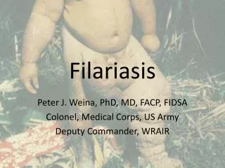
FILARIASIS M.PRASAD NAIDUMSc, (Medical) Ph.D. (Medical)
Filariasis (or philariasis) is a parasitic disease that is caused by thread-like roundworms belonging to the Filarioidea type. • These are spread by blood-feeding black flies and mosquitoes. • Eight known filarial nematodes use humans as their definitive hosts. These are divided into three groups according to the niche within the body they occupy.
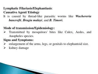
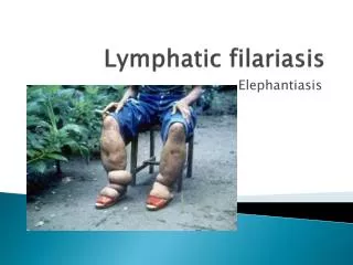
Lymphatic filariasis is caused by the worms Wuchereria bancrofti, Brugia malayi, and Brugia timori. • These worms occupy the lymphatic system, including the lymph nodes; in chronic cases, these worms lead to the disease elephantiasis.
The adult worms, which usually stay in one tissue, release early larval forms known as microfilariae into the host's bloodstream. These circulating microfilariae can be taken up with a blood meal by the arthropod vector; in the vector, they develop into infective larvae that can be transmitted to a new host.
Adult worm: Adult are minute,whitish thread like and are filariform in shape with smooth surface. Both the ends are tapering. Morphology
Individuals infected by filarial worms may be described as either "microfilaraemic" or "amicrofilaraemic", depending on whether microfilariae can be found in their peripheral blood. • Filariasis is diagnosed in microfilaraemic cases primarily through direct observation of microfilariae in the peripheral blood. • Occult filariasis is diagnosed in amicrofilaraemic cases based on clinical observations and, in some cases, by finding a circulating antigen in the blood.
Male worm measures 40mm in length and and 0.1 mm in breadth. Posterior end is sharply curved ventrally with two spicules of unequal length. Male worm
The female measures 80-100mm in length and are longer than males. The female are viviparous and lay eggs containing well developed embryos, the microfilariae. The microfilariae remain coiled together within the uterus and vagina of the mature gravid female worm. Female worm
The third stage larva is called as microfilaria. Found in peripheral blood and often in the hydrocele fluid and chylous urine. They are transparent and colourless by a hyaline sheath. The microfilaria can live up to 70 days in the human blood. Microfilaria
There is a marked periodicity in the circulation of microfilaria in the blood. They will be present in high numbers in the peripheral blood during a 4 hour period at mid night and scanty or absent at day time. This type of periodicity is called as nocturnal periodicity. The exact mechanism of periodicity is not clearly known.Body temperature,oxygen,sleeping habits etc may influence the periodic pattern of the microfilaria. Microfilarial periodicity
Third stage larva: It is the infective form of the filarial worm for humans. It is found only in mosquito vectors. The microfilaria are elongated, filariform and measure 1.4mm to 2m in length.
African green leaf monkeys. The adult worms are found in the sacral,para-aortic and inguinal lymph nodes and the thoracic duct and associated vessels. Laboratory animal:
Completes life cycle in two hosts. Definitive host: Man. Intermediate host: Mosquitoes: Culex quinquefaciatus, Anopheles Aedes Species. Life cycle:
The pathogenic effect is produced by adult worms of Wuchereria,living or dead. The infections are mainly classified into two forms 1.CLASSICAL FILARIASIS. 2.OCCULT FILARIASIS. Pathogenesis and clinical features
Causes • Human filarial nematode worms have complicated lifecycles, which primarily consists of five stages. After the male and female worms mate, the female gives birth to live microfilariae by the thousands. The microfilariae are taken up by the vector insect (intermediate host) during a blood meal
In the intermediate host, the microfilariae molt and develop into third-stage (infective) larvae. • Upon taking another blood meal, the vector insect injects the infectious larvae into the dermis layer of the skin. After about one year, the larvae molt through two more stages, maturing into the adult worms.
Filarial lymphangitis usually accompanied by a rise of temperature ranging from 1030 c to 104o F which may continue for several days. The temperature comes down with profuse sweating. The fever is associated with localised sign of inflammation of the lymphatic vessel where the adult worm lies. Examination of the blood may reveal microfilaria at this stage. Filarial fever:
The most spectacular symptom of lymphatic filariasis is elephantiasis—edema with thickening of the skin and underlying tissues—which was the first disease discovered to be transmitted by mosquito bites. • Elephantiasis results when the parasites lodge in the lymphatic system. • Elephantiasis affects mainly the lower extremities, while the ears, mucous membranes, and amputation stumps are affected less frequently.
However, different species of filarial worms tend to affect different parts of the body;Wuchereria bancrofti can affect the legs, arms, vulva, breasts, and scrotum (causing hydrocele formation), whileBrugia timori rarely affects the genitals.Those who develop the chronic stages of elephantiasis are usually amicrofilaraemic, and often have adverse immunological reactions to the microfilariae, as well as the adult worms.
The subcutaneous worms present with skin rashes, urticarial papules, and arthritis, as well as hyper- and hypopigmentation macules. Onchocerca volvulus manifests itself in the eyes, causing "river blindness" (onchocerciasis), one of the leading causes of blindness in the world. • Serous cavity filariasis presents with symptoms similar to subcutaneous filariasis, in addition to abdominal pain, because these worms are also deep-tissue dwellers.
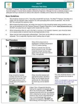
Filariasis is usually diagnosed by identifying microfilariae on Giemsa stained, thin and thick blood film smears, using the "gold standard" known as the finger prick test. The finger prick test draws blood from the capillaries of the finger tip; larger veins can be used for blood extraction, but strict windows of the time of day must be observed. Diagnosis:
Blood must be drawn at appropriate times, which reflect the feeding activities of the vector insects. Examples are W. bancrofti, whose vector is a mosquito; night is the preferred time for blood collection. Loa loa's vector is the deer fly; daytime collection is preferred. • This method of diagnosis is only relevant to microfilariae that use the blood as transport from the lungs to the skin.
Some filarial worms, such as M.streptocerca and O. volvulus, produce microfilarae that do not use the blood; they reside in the skin only. For these worms, diagnosis relies upon skin snips, and can be carried out at any time.
Polymerase chain reaction (PCR) and antigenic assays, which detect circulating filarial antigens, are also available for making the diagnosis. The latter are particularly useful in amicrofilaraemic cases. Spot tests for antigen are far more sensitive, and allow the test to be done any time, rather in the late hours.
Lymph node aspirate and chylus fluid may also yield microfilariae. Medical imaging, such as CT or MRI, may reveal "filarial dance sign" in chylus fluid; X-ray tests can show calcified adult worms in lymphatics. The DEC provocation test is performed to obtain satisfying numbers of parasites in daytime samples. Xenodiagnosis is now obsolete, and eosinophilia is a nonspecific primary sign.
The recommended treatment for people outside the United States is albendazole (a broad-spectrum anthelmintic) combined with ivermectin. A combination of diethylcarbamazine and albendazole is also effective. All of these treatments are microfilaricides; they have no effect on the adult worms. Different trials were made to use the known drug at its maximum capacity in absence of new drugs. Treatment
In a study from India, it has been shown that a formulation of albendazole has better anti-filarial efficacy than albendazole itself. • In 2003, the common antibiotic doxycycline was suggested for treating elephantiasis
Filarial parasites have symbiotic bacteria in the genus Wolbachia, which live inside the worm and seem to play a major role in both its reproduction and the development of the disease. • Clinical trials in June 2005 by the Liverpool School of Tropical Medicine reported an eight-week course almost completely eliminated microfilaraemia
1.Mosquito control: Physical:Mosquito net,effective drainage system. Chemical :Mosquito repellents,DDT Biological:Gambusiya fish. 2.Treatment of the patients Prevention and control