Download
1 / 48

E N D
RECOMBINANT DNA M.PRASAD NAIDU Msc Medical Biochemistry, Ph.D Research scholar.
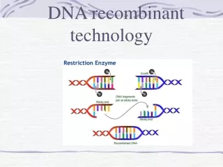
RECOMBINANT DNA The ability to cut and paste genetic material for further analysis is due to the discovery of enzymes called restriction endonucleases. These enzymes cleave double stranded DNA at specific sequences, most commonly formed by 4, 6 or 8 base pairs. Different RE recognize sequences that are either frequent or quite rare. They are useful for different applications. Ex. EcoRI SmaI
More than 3000 REs have been isolated from bacteria. Their function, in vivo, is to cleave and promote degradation of viral DNA inside the host bacterial cells. Bacterial DNA is protected from cleavage because it’s methylated.
Restriction nucleases can cleave the DNA leaving overhanging single stranded tails (sticky) or blunt ends.
Rare restriction nucleases , such as EcoRI, can be used for mapping of DNA . This analysis involves cutting the sample DNA and separate the fragments according to size by gel electrophoresis.
Agarose gel electrophoresis: it’s prepared as a 1-2% agarose in buffer and cast horizontally on a tray containing several wells, for each sample. The bigger the DNA fragment the slower it will migrate down the gel, when the current is applied. Since the DNA is charged negatively, the direction of the migration will be from the negative to the positive electrode. The DNA is visualized by addition of a dye (ethydium bromide)that intercalates between the DNA bases and is fluorescent. The intensity of fluorescence is proportional to the amount of DNA in the sample. Show example
Problem: 2% agarose gel, in 50 ml TE buffer Ingredients: agarose powder 100x TE buffer
Ethidium bromide is an intercalating dye, which means it inserts itself between the bases that are stacked in the center of the DNA helix. One ethidium bromide molecule binds to one base. As each dye molecule binds to the bases the helix is unwound to accommodate the strain from the dye. Closed circular DNA is constrained and cannot withstand as much twisting strain as can linear DNA, so circular DNA cannot bind as much dye as can linear DNA. Ethidium bromide can easily get into your cells. Human DNA is linear, and stains well. This means that it can get into your DNA and untwist it.
DNA mapping Eco RI : 1100 bp 500 bp 100 bp Bam H: 900 800 Eco RI + BamH: 700 500 400 100
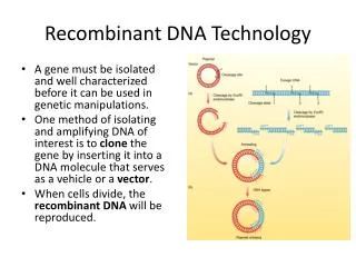
MOLECULAR CLONING The basic strategy in molecular cloning is to insert a DNA fragment of interest into a DNA molecule (called a vector) that is capable of independent replication in a host cell. The host cell is usually E. Coli, and the vector is a plasmid or a phage that can replicate producing million of progeny recombinant molecules. Plasmid or phage DNA can be isolated separately from the host genomic DNA and identified by cutting and sequencing.
Vectors for Recombinant DNA 1.Lambda (λ) phage. It’s used for either genomic or cDNA clones from eucaryotic cells.Sequences of DNA up to 15 kb can be inserted. -Insertion -packaging -E. Coli infection -Isolation of single clones
2. Plasmids. Smaller than phage, easier to manipulate, can replicate independently from the host cell. Plasmid DNA can be easily separated from the bacterial DNA and sequenced. 3. Cosmids and yeast artificial chromosome (YAC) are used To clone big pieces of genomic DNA (up to 45kb in cosmid and over hundreds of kb in YAC)
A plasmid vector is digested with EcoRI at a single site to produce two sticky ends. • A sample of human DNA is also digested with EcoRI to produce pieces with the same sticky ends. • Human DNA- or cDNA copied from mRNA using reverse transcriptase from retroviruses. • The two samples are mixed and allowed to hybridize, some molecules will form with pieces of human DNA inserted into the plasmid vector at the EcoRI site. • DNA ligase is used to covalently link the fragments. Creating Recombinant DNA
DNA denaturation: the two strands are separated by heat or chemical treatment. DNA/RNA hybridization: single stranded DNA or RNA is allowed to anneal to its complementary strand (either DNA or RNA) in controlled conditions (temperature and salt concentration).
How do we isolate large quantities of DNA for further characterization? Polymerase chain reaction (PCR)
The techniques was developed by Nobel laureate biochemist Kary Mullis in 1984 and is based on the discovery of the biological activity at high temperatures of DNA polymerases found in thermophiles (bacteria that live in hot springs). Most DNA polymerases work only at low temperatures. But at low temperatures, DNA is tightly coiled, so the polymerases don't stand much of a chance of getting at most parts of the molecules.
But these thermophilic DNA polymerases work at 100C, a temperature at which DNA is denatured. This thermophilic DNA polymerase is called Taq polymerase, named after Thermus aquaticus, the bacteria it is derived from. Taq polymerase, however, has no proofreading ability. Other thermally stable polymerases, such as Vent and Pfu, have been discovered to both work for PCR and to proofread.
We’ve got the sequence. What’s next. • Search databases to identify identical or similar sequences identified • by others and corresponding to known proteins. • You found a hit: you can give your sequence a name • Your sequence is novel: you characterize it
An increasing number of resources is available on the web to conduct searches. Ex. Sequence characterization (amino acid translation, presence of Intron/exon, promoter sequences, structural analysis of polypeptides, Cellular localization) Goal: to get clues about the identity or function of the candidate clone
You found a putative peptide open frame sequence. • Q. Is it a real protein? How can I test it. • Gene expression in procaryotes. The isolated DNA is cloned in a • vector containing a T7 promoter. Add amino acids, ATP generating • system, T7RNA polymerase, E. Coli extract (containing ribosomes and • enzymes for translation). • Run the product of the reaction on a acrylamide gel to identify the • protein.
Recombinant proteins can be also be expressed in yeast or in mammalian cells. Applications: studies of protein function in particular tissues or conditions (cancer).
Polymerase Chain Reaction (PCR) It allows to produce and isolate large amounts of single DNA molecules for which the complete or partial sequence is known. DNA Polymerase (Taq, Vent, or Pfu) F and R oligonucleotides Free deoxynucleotides Reaction buffer (includes Mg++) DNA Template (linear DNA, cDNA or genomic, plasmid, pure, fixed, from cells, etc.) DNA is amplified exponentially (1 copy 30 cycles = 1 billion copies)
PCR Variations: RT-PCR Real time PCR Degenerate primers PCR
RT-PCR: • Isolate RNA (total or polyA) • Convert to cDNA (complementary DNA, using the reverse transcriptase) • Use the DNA as template for the PCR reaction • Visualize fragment on agarose gel
Real time PCR It’s used for accurate quantitation of DNA samples. In real time PCR the concentration of a DNA sample is proportional to the amount of fluorescence generated at each round of amplification.
The real-time PCR system is based on the detection and quantitation of a fluorescent reporter. This signal increases in direct proportion to the amount of PCR product in a reaction. By recording the amount of fluorescence emission at each cycle, it is possible to monitor the PCR reaction during exponential phase where the first significant increase in the amount of PCR product correlates to the initial amount of target template.
The best method for quantitative detection of the amplicon uses fluorescent probes. The TaqMan probes use the fluorogenic 5' exonuclease activity of Taq polymerase to measure the amount of target sequences in cDNA samples. TaqMan probes are oligonucleotides that contain a fluorescent dye usually on the 5' base, and a quenching dye on the 3' base. When irradiated, the excited fluorescent dye transfers energy to the nearby quenching dye molecule rather than fluorescing (this is called FRET = Förster or fluorescence resonance energy transfer). Thus, the close proximity of the reporter and quencher prevents emission of any fluorescence while the probe is intact.
About | People | DNA Sequencing | Oligo Synthesis | Microarray | Real-Time PCR | ABI-Freezer Program | Seqweb
Degenerate PCRDegenerate Primers - What are they?Primers which have a number of options at several positions in the sequence to allow annealing to and amplification of a variety of related sequences.eg:5’-TCG AAT TCI CCY AAY TGR CCN T-3’Y = pYrimidines = C / T (degeneracy = 2X)R = puRines = A / G (degeneracy = 2X)I = Inosine = C / G / A / T N = Nucleotide = C / G / A / T (degeneracy = 4X) • Why... use degenerate primers? • to amplify (fish out) conserved sequences of a gene or genes from the genome of an organism. • to get the nucleotide sequence after having sequenced some amino acids from a protein of interest
Detection of nucleic acids Based on the principle of nucleic acid hybridization: Southern blot (DNA) Northern blot (RNA) In situ hybridization (intact chromosomes, cells, tissue slices, or embryos).
Southern Blot A DNA probe is hybridized to genomic DNA or cDNA. The DNA probe is labeled (radioactive, fluorescent, or chemoluminiscent). Southern blotting was named after Edward M. Southern who developed this procedure at Edinburgh University in the 1970s. DNA molecules are transferred from an agarose gel onto a membrane. Southern blotting is designed to locate a particular sequence of DNA within a complex mixture. For example, Southern Blotting could be used to locate a particular gene within an entire genome.
Digest the DNA with an appropriate restriction enzyme. • 2. Run the digest on an agarose gel. • 3. Denature the DNA (usually while it is still on the gel).For example, soak it in about 0.5M NaOH, which would separate double-stranded DNA into single-stranded DNA. Only ssDNA can transfer.
5. Probe the membrane with labeled ssDNA. This is also known as hybridization.Whatever you call it, this process relies on the ssDNA hybridizing (annealing) to the DNA on the membrane due to the binding of complementary strands. • Probing is often done with 32P labeled ATP, biotin/streptavidin or a bioluminescent probe.
6.Visualize your radioactively labeled target sequence. If you used a radiolabeled 32P probe, then you would visualize by autoradiograph. Biotin/streptavidin detection is done by colorimetric methods, and bioluminescent visualization uses luminescence.
Northern Blot Total RNA or poly(A) RNA is isolated on agarose gel, transferred to a nylon membrane and hybridized to a DNA probe. Usually, the labeled DNA probe is a region of a gene for which you want to study the expression pattern. Example: Is the estrogen receptor expressed in all tissues at the same level?
In situ hybridization: The probe is hybridized to either RNA or DNA on a slice of tissue, or cells in culture. The fluorescent microscope is used to visualize the bound probe. Example : in which tissue of the developing embryo is protein X first expressed?
Detection of proteins Antibodies (polyclonal and monoclonal) Western blotting: - protein extract - primary antibody (mouse or rabbit) - secondary antibody (labeled, goat IgG anti mouse or rabbit) -autoradiography
Gene function Gene transfer in plants and animals. DNA transfection : transient or stable Methods: calcium phosphate precipitation liposomes electroporation retrovirus Transgenic animals
Microinjection of DNA into a pro-nucleus • of a fertilized mouse egg. • Eggs are transfected to foster mothers and are allowed to develop • Some of the offspring will have the injected DNA incorporated • in their genome. • 2. Embryonal stem cells are derived from blastocysts. • Foreign DNA is transfected into ES cells. Transformed cells • are injected into blastocysts, which are transferred to foster • mothers. Chimera offspring is produced, mated to normal male, • transgene is incorporated in the offspring.
Plant viruses and plasmids Ti plasmid (from the Agrobacterium tumifaciens). The bacterium attaches to the leaves of plants and the Ti is transferred into plant cells where it becomes incorporated into chromosomal DNA. So vectors developed from Ti plasmids can be used as means of introducing recombinant DNA into sensitive plant cells.
In vitro mutagenesis Single point mutations Deletions Duplications Insertions Reverse genetics
Homologous recombination of a DNA molecule with its chromosomal copy. A mutated DNA can be integrated into the genome. The target gene can be inactivated, so the function can be identified. Antisense nucleic acids (RNA or single stranded DNA) Dominant inhibitory mutants. Mutant proteins can be introduced into cells by gene transfer and used to study the effects of blocking normal gene function (embryonic development in xenopus)