Download
1 / 46
460 likes | 474 Vues
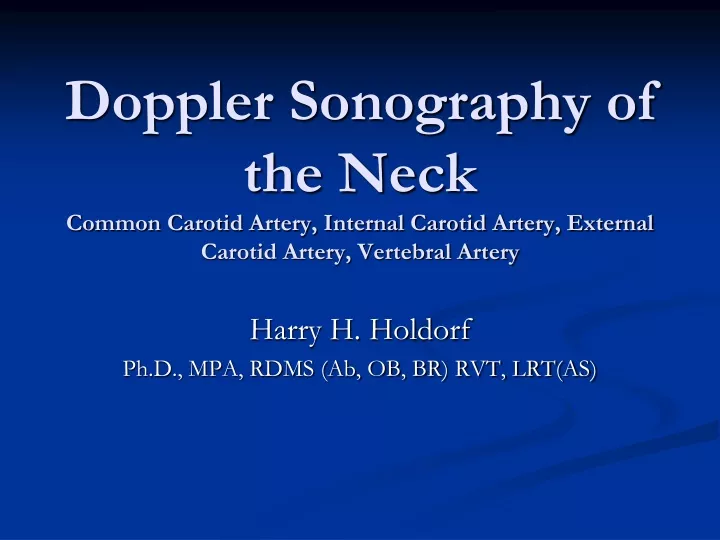
Doppler Sonography of the Neck Common Carotid Artery, Internal Carotid Artery, External Carotid Artery, Vertebral Artery. Harry H. Holdorf Ph.D., MPA, RDMS (Ab, OB, BR) RVT, LRT(AS). Table of Contents. Anatomy Indications for Ultrasound The Ultrasound examination 2-D Color Doppler

E N D
Doppler Sonography of the NeckCommon Carotid Artery, Internal Carotid Artery, External Carotid Artery, Vertebral Artery Harry H. Holdorf Ph.D., MPA, RDMS (Ab, OB, BR) RVT, LRT(AS)
Table of Contents • Anatomy • Indications for Ultrasound • The Ultrasound examination • 2-D • Color Doppler • Spectral Doppler • Diagnostic Criteria
Indications for Ultrasound • Evaluation of patients with TIA • Transient Ischemic Attack • Evaluation of patients with CVA • Cerebrovascular Accident • Evaluation of carotid bruits • Follow up of known disease • Monitor endarterectomy results • Evaluation of pulsatile neck mass
Carotid Ultrasound Examination • Patient supine • Neck slightly extended • Head slightly turned away from side being scanned • Linear transducer, 5-12 MHz • Protocols vary between operator, institution • B-mode, color Doppler, pulsed Doppler
Carotid Ultrasound Examination • B-mode images – Transverse/Sagittal • Images from proximal CCA to bifurcation • Bulb – widening of CCA near bifurcation • Bifurcation – identify and ICA and ECA • Color/Doppler • Proximal/Mid/Distal CCA • Bifurcation (color) • Prox/Mid/Distal ICA • ECA • Vertebral • More images for pathology
Anatomy • Aortic Arch • Innominate or Brachiocephalic artery • Right subclavian artery • Right common carotid • Left common carotid artery • Left subclavian artery • CCAs lie • posterolateral to thyroid • Deep to sternocleidomastoid muscle
Anatomy • Right CCA originates from innominate artery • Left CCA originates from aortic arch • CCAs divide into ECA and ICA • ICA has no branching vessels in the neck; may have slight dilation just past its origin; supplies face and head; lies posterior in the neck • ECA has branching vessels; usually smaller than ICA • Vertebral Arteries – lie between transverse process of the vertebrae
Answers to previous slide Aortic arch angiogram 1. Innominate art. 2. Right subclavian art. 3. Right common carotid art. 4. Right vertebral art. 5. Left common carotid art. 6. Left vertebral art. 7. Left subclavian art.
Deep dissection of neck.BIF = bifurcation,SCM= sternocleidomastoid,TM= trapezius muscle
ICA The internal carotid artery supplies the brain, the eye, the forehead, and part of the nose. It enters the cranial cavity via the carotid canal in the temporal bone. It gives off no branches within the neck region. The internal carotid artery supplies perfusion to the anterior and middle portion of the brain, the eye and its appendages and sends branches off to the forehead and nose. It originates at the carotid bifurcation and courses, via a number of curvatures, through the base of the skull into the Circle of Willis. The proximal-most portion of this vessel is rather bad bulbous and is referred to as the bulb. Distal to the bulb, the artery narrows and is referred to as the ICA proper. Anatomically, the ICA is distinct from the ECA in that there are no branches until it passes through the base of the skull. After coursing anteriorly to the transverse processes of the first three cervical vertebrae (C1 –C3), it enters the carotid canal of the petrous portion of the temporal bone. The intra-petrous portion of the ICA is also referred to as the cavernous portion of the ICA. After coursing through the base of the skull, its first branch arises, the ophthalmic artery, which supplies blood to the retina. (This is of clinical importance when discussing amaurosisfugax). At the base of the brain, the ICA gives rise to the anterior and middle cerebral arteries. The site of this branching is often referred to as the carotid siphon.
External carotid artery. Superior thyroidal and lingual artery branches are seen in the submandibular space. The superficial temporal artery lies anterior to the ear.
Vertebral Artery • Subclavian Steal • When a stenosis or occlusion of a the proximal subclavian or innominate and the effected side “steals” blood from retrograde vertebral artery flow
Distinguishing characteristics of the vertebral artery: First branch off subclavian Enters foraminal canal at C6 Asymmetrical size: left > right Low resistance flow
Carotid Ultrasound Examination • Differentiate ICA/ECA • ICA is usually lateral • ICA usually larger than ECA • ECA has branches • Temporal tap – tapping of superficial temporal artery in the pre-auricular area; will appear as “saw-teeth” on spectral waveform
Carotid Ultrasound ExaminationGray Scale Image • Gray-Scale Image • Vessel Wall Thickness • IM Complex - .8 mm may be considered abnormal • Plaque Characterization • Homogeneous – smooth surface, uniform echogenicity (p 953) • Calcified – echogenic with posterior shadowing (p 954) • Heterogeneous – complex with at least one sonolucent area corresponding to at least 50% of plaque volume (p 955) • Interplaque hemorrhage – “Swiss cheese” appearance • Plaque ulceration – focal depression/break in surface; or anechoic area that extends to plaque surface; use color/power Doppler (p 952) • Some soft plaque may be very difficult to see on US • Evaluation of Stenosis – % stenosis by diameter/area – taken in transverse plane (p 957)
ICA: Duplex characteristics of the ICA. Lowresistance waveform. Here a clear spectral window isseen under the spectral envelope.
Carotid Ultrasound ExaminationColor Doppler • Color assignment depends on direction of flow relative to the transducer • Color saturation indicates variable velocities • Lower velocities depicted toward baseline, usually deeper shades • Higher velocities usually brighter shades/colors • Tagging of high velocities • Gain/Scale settings
Duplex characteristics of the ECA . Rapidsystolic upsweep with high resistance flowpattern during diastole. “Temporal tapping” canbe seen as spectral oscillations during diastole.
Carotid Ultrasound Examination • Altered flow patterns • Pathology • Normal • At area where vessel branches • Where diameter of vessel changes • Tortuosity • High flow jets after stenosis • Low trickle flow
Carotid Ultrasound EvaluationSpectral Analysis • Graphic display of velocities and direction of moving red blood cells in the sample volume • X axis, Y axis • Direction of flow • Amplitude of “velocity component” (# of RBCs within each velocity component = brightness of the trace) • Spectral window – black zone between baseline and spectral line
Carotid Interpretation • Spectral Broadening – occurs when blood cells move with a wider range of velocities; normal spectral window will be filled in • Increases in proportion to the severity of stenosis • Do not confuse with over gain or vessel wall motion • See Rumack p 960 Fig 25-18
Carotid Interpretation • Doppler cursor should be in the center of the vessel, parallel to the vessel walls; on high velocity jets • Doppler angles 60 degrees or less • Doppler samples obtained just proximal to, at and just beyond stenosis • 1 cm intervals distal to plaque
Carotid Ultrasound • ECA – feeds high resistance vascular bed; velocity rises sharply during systole, falls quickly during diastole • ICA – feeds lower resistance brain circulation; large quantities of flow continue during diastole; supplies brain; velocities usually increase from prox to distal • CCA – resembles ICA waveform, with diastolic flow above the baseline
Carotid Ultrasound ExaminationDiagnostic Criteria • Carotid artery stenosis (%)– reduction in diameter • Carotid artery occlusion • No flow detected with color, pulsed Doppler • Thrombus/plaque fills vessel lumen • Reversal of flow proximal to occluded segment • Plaque characterization – as discussed above • Numerical values of some diagnostic criteria may very between institutions • See Rumack p 960
Carotid Ultrasound EvaluationDiagnostic Criteria • Degree of stenosis assessed using • ICA pre-systolic velocity • ICA End Diastolic velocity • PS ICA/CCA ratio • PED ICA/CCA ratio • No established criteria for grading ECA stenosis - occlusive plaque in ECA is less common and rarely clinically significant (Rumack) • Velocity criteria used to grade CCA stenosis have not been well established (Rumack) • See Rumack p 967
Carotid Ultrasound • > 50 % - mild stenosis; not hemodynamically significant • 50 - 69% moderate stenosis; follow up for progression; hemodynamically significant • 70 – 99% - severe stenosis; operable lesion • Occlusion – concern regarding hemodynamic status of intracranial circulation, not an operable lesion • Ulceration – considered clinically significant; can rupture or hemorrhaging; plaque can continue to embolize or occlude ICA
Superficial temporal artery: Terminal branch of ECA. Supplies skin over frontal and temporal regions of scalp. The superior temporal artery, which can be palpated just anterior to the pinna of the ear, is sometimes examined using duplex ultrasound in patients with suspected temporal arteritis. TEMPORAL TAP METHOD
Circle of Willis (connecting the two) Major collateral pathway of brain Basilar artery formed from fusion of both vertebrals Internal carotid arteries Anterior cerebral Middle cerebral Basilar artery Posterior cerebral Communicating vessels Anterior communicating artery (acoa) Posterior communicating artery (pcoa)
References • Araki, C.T. Introduction to Non-Invasive Vascular Testing. UMDNJ Vascular Technology Program. • Rumack C., Wilson, S. & Carboneau. Diagnostic Ultrasound, 4thEdition, Vol 1. 2005. • Image slide 4 retrieved on November 27th, 2012 from http://www.vascularultrasound.net/vascular-anatomy/arteries/extracranial • Image slide 6 retrieved on November 27th 2012 from http://www.medison.ru/uzi/eho41.htm • Image slide 7 rwetrieved on November 27 2012 from http://www.specialistvascularclinic.com.au/carotid-interventions.html • Image slide 8 retrieved on November 27th from http://www.medicern.co.uk/products/Colour-Portable-Ultrasound/M7-Colour-Doppler-Portable-Ultrasound.html • Image slide 10 retrieved on November 27th from http://www.ultrasoundpaedia.com/normal-carotids/ • Image slide 24 retrieved on November 28, 2012 from http://www.ajronline.org/content/177/1/53.figures-only