Download
1 / 29
300 likes | 581 Vues
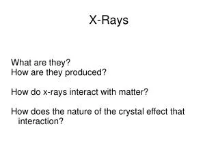
X- Rays. X-rays are generated from the interaction of accelerated e - ’s & a target metal (tungsten) Patient is placed between X-ray tube and silver halide film X-rays passed through the body are absorbed in direct proportion to tissue density

E N D
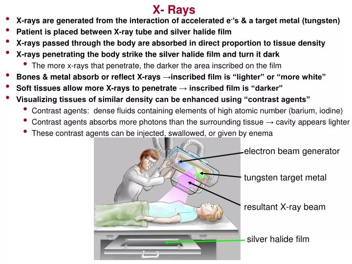
X- Rays • X-rays are generated from the interaction of accelerated e-’s & a target metal (tungsten) • Patient is placed between X-ray tube and silver halide film • X-rays passed through the body are absorbed in direct proportion to tissue density • X-rays penetrating the body strike the silver halide film and turn it dark • The more x-rays that penetrate, the darker the area inscribed on the film • Bones & metal absorb or reflect X-rays →inscribed film is “lighter” or “more white” • Soft tissues allow more X-rays to penetrate → inscribed film is “darker” • Visualizing tissues of similar density can be enhanced using “contrast agents” • Contrast agents: dense fluids containing elements of high atomic number (barium, iodine) • Contrast agents absorbs more photons than the surrounding tissue→cavity appears lighter • These contrast agents can be injected, swallowed, or given by enema electron beam generator tungsten target metal resultant X-ray beam silver halide film
X-ray View of a Gunshot Wound (Bullet has split into fragments)
X-Ray Mammography Breast Cancer ! Normal Breast Breast with Cysts and Fibrotic Changes
Classic X-ray view of “Lung Infiltrates” caused by Pneumonia. Notice the increased “whiteness” close to the sternum
X-ray view of broken ribs in an infant …. ……caused by child abuse. Specifically, by holding the baby by the chest and shaking him violently.
Computed Tomography (“CT Scan” or “Cat Scan”) • The scanner device incorporates a moving table & a revolving X-ray tube • The table moves the patient back and forth through the revolving X-ray emissions • The X-ray emitter moves (revolves) in a 360o arc around the patient • Instead of film, the CT scanner collects emitted X-rays via a collector • This collector is called a SCINTILLATOR • Scintillator transforms X-ray’s into a proportionally strong electric current • The electric current is then converted into a number of images (“slices”) • Contrast dyes may be used for image enhancement • Tool of choice for most stroke cases
X-ray collector bank rotates around patient CT scan X-ray tube
CT scan color enhancement Purple area denotes destruction of normal brain tissue which is colored green
Magnetic nuclei are abundant in the human body (H,C,Na,P,K) and spin randomly • Since most of the body is H2O, the Hydrogen nucleus is especially prevalent • Patient is placed in a static magnetic field • Magnetized protons (spinning H nuclei) in the patient align in this field like compass needles • Radio frequency (RF) pulses then bombard the magnitized nuclei causing them to flip around • The nuclei absorb the RF energy and enter an excited state • When the magnet is turned off, excited nuclei return to normal state & give off RF energy • The energy given off reflect the number of protons in a “slice” of tissue • Different tissues absorb & give off different amounts of RF energy (different resonances) • The RF energy given off is picked up by the receiver coil & transformed into images • MRI offers the greatest “contrast” in tissue imaging technology (knee, ankle diagnosis) • cost: about $1450 - $2000 • time: 30 minutes - 2 hours, depending on the type of study being done Magnetic Resonance Imaging Closed (traditional) MRI scanner Open MRI
Magnetic Resonance Imagingtissues composition & signal intensity Tissue Signal Intensity T1 Signal Intensity T2 Fat high (whitish) intermediate Muscle intermediate (gray) intermediate Hyaline Cartilage intermediate intermediate - low (dull gray) Ligaments & Tendons low (dark gray) low Cortical Bone low low Granulation Tissue intermediate high Fibrous Tissue low low Hemorrhage / Edema high - intermediate high Immature Scar intermediate - low low to high Mature Scar low low
Significant meniscus tears (indicated by the green arrows) in frontal (left) and the sagital (below) planes
Grade 3 ACL tear (note “lighter” region where the “darker” region used to be. This indicates tissue disruption and associated fluid buildup) Normal ACL (note “darker” region indicating normality)
Bone Scan • Measures the rate of bone formation • Any disease that injures bone will cause new bone to form • This process is a very sensitive measure of bone disease processes • Often used for detecting cancer mets (breast, prostate), fractures, & infection • Can be used to detect avascular necrosis of bone • Procedure is done by injecting a technetium labeled phosphate (radioactive) • Pictures are taken using a gamma camera…… • Immediately after injection, 3 hours post injection, & 24 hour post injection • Dose of radiation is small • Takes about an hour to complete
Bone Scan of Non-malignant Osteoid Bone Tumor “hot spot” indicating ↑uptake of isotope in right femur
Bone Scan of Prostate Caner Metastases are indicated by the Green arrows
Positron Emission Tomography (PET) Scan • Device measures metabolism via the decay of radioactive tracers in tissues with higher than normal metabolic activity (such as cancer) • Patient is injected with FluorDeoxyGlucose • Glucose bound to Fluorine 18 (radioactive) • Diseased organs & tissues process FDG at a higher rate than normal tissues making FDG concentration higher in diseased tissue • Positrons are emitted by FDG and collide with electrons, emitting γ radiation • Radiation picked up by γcamera • Computer reconstructs the radioactivity into 3 dimensional images of organ or area • with higher than normal FDP uptake • Procedure performed as outpatient • Takes about 2 hours • Results available to physician within 48 hours
PET Scan showing Non Hodgkins Lymphoma (Green Arrows) before & after 6 months of chemotherapy
Dual Energy X-ray Absoprtometry (DXA or DEXA) • Used to test for bone mineral density (BMD) ie. Osteoperosis • Thin X-ray beam is passed through the hip and lower spine regions • Computer calculates how much X-ray energy is absorbed by the bones • Computer compares results with an average 20 year old (T-score) and an average age, race and gender peer (Z-score) • Results are plotted on a norm graph and given to the radiologist
Dual Energy X-ray Absoprtometry (DXA or DEXA) GE LUNAR Prodigy DEXA...in the Applied Exercise Science Lab
Color enhanced DEXA Scan: T-score: -1.8 The more dense regions are red/orange/yellow