Download
1 / 66
670 likes | 995 Vues
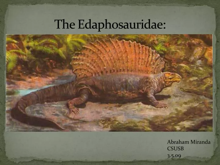
The Edaphosauridae :. Abraham Miranda CSUSB 3.5.09. Edaphosauridae. Barrel bodied Small heads Originally lumped together, the subclass Edaphosauria include Edaphosauridae Casea Lupeosauridae Nitrosauridae

E N D
The Edaphosauridae: Abraham Miranda CSUSB 3.5.09
Edaphosauridae • Barrel bodied • Small heads • Originally lumped together, the subclass Edaphosauria include • Edaphosauridae • Casea • Lupeosauridae • Nitrosauridae • Romer appeared to have decided Edaphosaurus should have their own subclass, but he was overruled • Romer compared the Edaphosaurus to Casea because of their pelycosaur similarities
Commonalitys of the Edaphosauridae • Projection off the frontal bone, creating a large lappet • Quadratojugal not connected to the subtemporal bar • Lacrimal bone reaches from the orbits to the naris • A narrow skull table • Have a supraorbial brow or shelf created from the prefrontal, frontal and post frontal bones
The Edaphosaurid head • Postorbital process reduced • No connection with the postorbital bone and supratemporal bone • Temporal bar is created from the jugal and squamosal bones • Quadratojugal jugal is reduced
Back of the head- Occiput • Supraoccipital is smaller compared to sphenacodonts
Jaw and Tooth • Articulation of the jaw joint is below the rows of teeth • Dentary bone has a big coronid process • The prearticular bone is rotated to fit under the pterygoid process of articular bone • All marginal teeth slightly enlarged at distal portions
The Axial skeleton and the sail • Sail: Formed by the neuro spinous processes of the cervical, thoracic, and lumbar vertebrae • Neural spines are circular at distal ends • Neural spines are laterally compressed at proximal ends • Cervical neural spines bend anterioly • Posterior neural spines bend posteriorly • Neural spine tubercles paired at the proximal ends
The Neurospine process Sail • Neural spines enlongated on presacral bones • Short transverse tubercles • Distal neural spines loose dual cylinder structure • Stolen from Adam Huttenlocker
Axial and Appendicular skeleton • Limbs are short • All Edaphosaurs have a curve to ribs • Tubercular head of ribs are small ridged
Ianthasaurus • Oldest and most primitive of the Edaphosauridae • Two well-preserved specimens found in Garnett, Kansas • First found in the Upper Pennsylvanian Rock Lake Shale of the Stanton formation. • A stream valley in which there was a slow transgression of flood waters. • First unearthed by P.E. Peabody in 1957, and thought to be a Dimetrodon, Reisz in 1982 confirmed this unknown specimen as an edaphosaur • Maintained edaphosaur spines, but did not really fit in with the rest of the genus, so a new genus was erected: Ianthasaurus • Only known species of Ianthasaurus: Ianthasaurushardestii • Insectivore
Ianthasaurushardestii • Pre and Postfrontal bones articulate to the Parietal • Reduced quadratojugal from the temporal bar • Small temporal fenestra • Long and low maxilla
Tooth and jaw • 27-29 teeth • Enlongate maxilla • Caniniform teeth • Sharp and recurved posteriorly dentition • Homodont dentition • No toothplates • Palatine teeth infer insectivory
Cervical Vertebrae • Centrum lengths of the Cervical vertebrae are greated than the rest of the presacral vertebrae • Neural spines running of the cervical vertebrae are thicker and more robust
Dorsal Vertebrae with ribs • Transverse processes of the vertebrae are short • Ribs are not strongly curved like the rest of the edaphosaurs • Longest neural spine is at vertebrae 17
Lumbar vertebrae • 29 Presacral vertebrae • Lack tubercles on the neural spines
Neural Spine Sail • Neural spine sail is the smallest of the Edaphosauridae • No more than 5 tubercles on neural spines • 27 Neural spines in the sail • No central elements in all neural spines • Neural spines lean forward in cervical region and rearward in the lumbar region • Neural spines begin at third cervical vertebrae on down to the second to last lumbar vertebrae • Measurements of the centra impossible to crush and bad preparation • Proximal portions of the neural spines are laterally compressed • Neural spines are subcircular at the basal tubercle • Considered a tool for heat exchange over a sexual dimorphism or “solar collector”
Appendicular Skeleton • Right scapulocorocoid • Left pelvis • Partial manus • Left humerous • Only 2 usable Ianthasaurus have been recovered
Scapula, corocoid, Humerous • Medial view: the scapular and anterior corocoid is exposed. • Posterior corocoid is unavalible • Scapula and corocoid are separated by nature or by degradation along suture contact • Gleniod is too poorly preserved • Supragleniod foramen exist? • At the anterior margin of scapular blade, a notch is seen in many pelcosaurs • Function? • Anterior corocoid expanded dorsoventrally and with a convex shape. • Function?
The Humerus • Humerus badly preserved and featureless • Distal end of humerous is narrow and no supinator process seen • Diaphysis is almost perfectly round in cross section • No evidence of the epiphyses being twisted about the bone
Manus • Identified as the ulnare, intermedium medial and lateral centrale • Nothing can be offered, not enough.
Left Pelvis • Illuim is well-developed with a blade-like processes extending posteriorly • The anterodorsal process is smaller than the posteriordorsal process
Pelvis,Ribs • Ribs are slighly curved with a smooth tubercular bump • Complete ilium has been found • Pubis and ischium have been lost • Iliac blade is extended posteriorly like other primitive pelycosaurs • Obturator foramen on posterior edge , but posterior boarder is opened • Indicates that this specimen is immature • Ischium retains the structure of other early pelycosaurs • Pubis and ischium are narrowly connected below the acetabulum
Glaucosaurus • Lived in the Lower Permian of North-Central Texas • Represented by a single skull • Shares 5 synapomorphies with Edaphosaurs
Glaucosaurus What is G? G= 5 reasons
5 Reasons to be an Edaphosaur • Transverse flange of pterygiod is missing
5 Reasons to be an Edaphosaur • No caniniform teeth • No caniniform region • Premaxillary and maxillary teeth identical
Edaphosaurus • 8 species currently recognized • Edaphosaurus • E. boanerges • E. cruciger- largest sail • E. pogonias • E. novomexicanus • E. colohistion • E. credneri • E. raymondi
Recovered Edaphosaur Remains • Most species are described by 1 or 2 badly preserved skulls • Many specimens found in the Geraldine Bonebed in Archer County, Texas • Larger barrel shaped body over Ianthasaurus
Edaphosaurus boanerges and friends • Dozens of specimens found in North-Central Texas • Many complete skulls • No so many complete skeletons • Used as the model for all Edaphosaurs
Skull information • Reduced skull size • Head is the size of 5 dorsal centra • Not as enlongate as the Ianthasaurus • Process of the postorbital is short, not extending to the parietal foramen • Nasal bone is ¾ the size of the frontal • Subtemporal bar is displaced superiorly • Temporal fenestra is enlarged anteriorly and posteriorly
Supraorbital shelf is wide and deep concealing the orbits • Lacrimal bone is thin at the maxillae and progressively thickens posteriorly suggesting it carries some kind of load and transfers it to the prefrontal • Prefrontal and lacrimal form a buttress attachment for reinforcement • The nasal bone has a “scarred shelf” and sutures that lock into the prefrontal bone as well as thicken around the orbit. • Suggests that carries load for feeding
Edaphosaurs • Premaxilla is enlongated in cross section • Premaxilla has 5 teeth, but none have survived to determine dimensions • The maxilla accomodates • 18-21 teeth • No Caniniform teeth • No Caniniform region • No pterygiod flange • All Homodont/isodonty dentition
Ventral and medial aspects • Jaw is suspended way below the upper tooth row • Well developed tooth plates on palate and mandible • Densely packet teeth • Tooth plates form on the palate and inner aspect of the mandible • Palatal plate consists of the pterygoidectopterygiod, and palatine
Ventral and medial aspects • Mandibular tooth plate is formed by the coroniod, posterior coroniod, and prearticular bones • Denticulated plate is formed by the ant coroniod, coroniod and prearticular bones • 120-150 teeth per palatal bone plate • Maxilla bone is twisted out laterally • Dentary bone is twisted in medially • Purpose?
Edaphosaur mandibles • Massive tooth plate found on the mandible, bigger than on the maxilla/palate • Deeply cut through mandibularsymphysis • Jaw articulation denoted propalinal or front to back movement of jaws • Maxillary teeth progressively angle backwards as you go to the dentary bone
Edaphosaur Teeth • Reduced homodont dentition • Isodonty in the marginal teeth cropped plant matter • Teeth are distally swollen • Fine serrated tips curving backwards • Palatal and mandibular tooth plates served as primary grinders of plant matter • Posterior maxillae and dentary teeth assisted with minor grinding
Occipital view of the skull • Supraoccipital has little lateral exposure • Tabular bones are thick towards the parietal, but they are thin and suture into paraocciptal bones • Postorbital is a slender sigmiodal bone. .
Cervical Vertebrae • Cervical vertebrae are extremely short compared to the dorsal and lumbar vertebrae • Ianthasaurus has longer centra on their cervical vertebrae • Contain neural spines that are enlongated with longitudinal ridges at the sides of the spines
Dorsal Vertebrae • Anterior to mid dorsal neural spines are tall and pointed. E. boanerges has the second longest neural spines • Neural spines are tall and pointed with a slight posterior angulations • Multiple lateral tubercules that are arranged laterally across the neural spines
Sacral and Caudal Vertebrae • Neural spine tips of sacral and caudal vertebrae are roughened or crenulated like a castle • They have longitudianal ridges that are rough Anterior view w/ R rib