Download
1 / 41
410 likes | 639 Vues
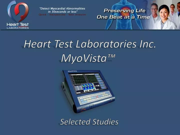
Heart Test Laboratories Inc. MyoVista™. Selected Studies. About Heart test Laboratories. HTL is a Texas based Company HTL was established in 2007 HTL has developed and released its“ patent pending” MyoVista™, a portable diagnostic device for the screening of Myocardial Ischemia (MI).

E N D
Heart Test Laboratories Inc. MyoVista™ Selected Studies
About Heart test Laboratories • HTL is a Texas based Company • HTL was established in 2007 • HTL has developed and released its“patent pending” MyoVista™, a portable diagnostic device for the screening of Myocardial Ischemia (MI)
About Beaufort Medical • Beaufort Medical is a Dubai based Company • Beaufort Medical was established in 2005 • Beaufort Medical provides a range of services including the distribution of medical devices in the MENA region including the distribution of the MyoVista™ Diagnostic Device
US STUDY MIECG - 2008 • MIECG (Myocardial Ischemia Electrocardiogram) A device that acquires and analyzes ECG waveforms in order to predict ischemia in patients that do not have symptoms and or are missed by conventional clinical pathways.
US STUDY MIECG – 2008 (Cont’d) • Premise: ECGs can be acquired and analyzed in an outpatient setting and can be predictive of ischemia: therefore leading to early identification of silent ischemia which will reduce the incidences of myocardial infarction due to unidentified patients with existing myocardial ischemia.
US STUDY MIECG – 2008 (Cont’d) • Objectives: The aim of this study was to acquire ECG data from a small population in the United States and compare the data to a 300 patient study performed in China. Data from China claimed above 90% predictive accuracy for the ischemic. The small US comparative study was performed at the Avista Adventist Hospital cardiac cathlab located in Louisville, Colorado.
US STUDY MIECG – 2008 (Cont’d) • Study Findings: • Correlation of test results to angiographic findings: • Traditional 12 lead ECG = 61% • Chest Pain with the emergent patient = 48% • Stress testing= 75% • MIECG = 87% (Highest)
US STUDY MIECG – 2008 (Cont’d) • Percentage of correlation with angiography
US STUDY MIECG – 2008 (Cont’d) • Percentage of False Positive Results
US STUDY MIECG – 2008 (Cont’d) • Percentage of False Negative Results
ASE 2011 • Title: PI-51 - Myocardial Stretch Related Repolarization and Relaxation Abnormalities of the Left Ventricle in Systemic Hypertension • Authors: Giuseppe Caracciolo, Shantanu Sengupta, Haruhiko Abe, Jagat Narula, Partho P. Sengupta. University of California, Irvine, Orange, CA
ASE 2011 (Cont’d) • Background: There is limited evidence to link myocardial stretch with the electrical and mechanical changes of the left ventricle (LV) in clinical settings. We hypothesized that increased systolic segmental stretch and cardiac repolarization abnormalities in patients with systemic hypertension result in dyssynchronous onset of LV segmental relaxation.
ASE 2011 (Cont’d) • Methods: Myocardial repolarization was assessed by signal processed electrocardiographic potentials (iECG, MyoVista in relation to 2-dimensional LV normal strains. We also obtained regional time volume curves by 3-dimensional speckle tracking echo cardiograph. Segmental dyssynchrony in onset of LV relaxation was measured as the standard deviation of the cross-over point of minimum systolic volume for the number of segments analyzed.
ASE 2011 (Cont’d) • Conclusions: LV relaxation abnormalities in patients with systemic hypertension are characterized by abnormal systolic stretch in longitudinal direction and repolarization abnormalities which account for dyssynchronous onset of LV
Angiogram Before & After • Facility: Fu Dan University Teaching Hospital Cardiology Department, Shanghai, China. • Professor: Yu Hong, MD , test date: Jan. 06, 2012 • These tests were taken within a Catheterization Lab. These slides show the test data of before and after results of an angiogram.
Angiogram Before & After • Facility: Fu Dan University Teaching Hospital Cardiology Department, Shanghai, China. • Professor: Yu Hong, MD , test date: Jan. 06, 2012 • These tests were taken within a Catheterization Lab. These slides show the test data of before and after results of an angiogram.
Angiogram Before & After (Cont’d) • Conclusion: There are no significant changes within the before and after ECG waveform test results. • The MyoVista 2D atlas waveform shows significant obvious changes in the before and after test results. • The MyoVista automatic diagnosis contains very readable test results information; this allows for easy analysis of the patient data.
Angiogram Before & After (Cont’d) • Conclusion (Cont’d): The MyoVista icon diagnosis displays simple targeting of the problematic areas. • The numerical index shows significant quantitative data of the patient's nature heart interactions. • numerical index shows significant quantitative data of the patient's nature heart interactions.
Angiogram Before & After (Cont’d) • Conclusion (Cont’d): Due to these results, MyoVista can be used for multiple different cardiology functions such as screening, clinical functions in the ER, in the OR, in ICU, in the CCU, and an drug treatment assessment of patient, etc. Therefore, this is a pivotal invention in the development of clinical diagnosis and basic cardiac assignment for many kinds of future patients.
Angiogram Before & After (Cont’d) • The following slides show the many differences in the MyoVista data before and after these procedures.
Patient # 4 • The patient was enrolled in the Heart Test Laboratories clinical trial at the Italian Medical Center in Asuncion, Paraguay. • Patients were put on a standard FDA approved 12-lead ECG device (the Nihon Khoden), then the MyoVista device, and then a coronary angiogram.
Patient # 4 (Cont’d) • Patient # 4 displayed a “normal” ECG reading, a severe “abnormal” i-ECG (the MyoVista informatics Electrocardiography) reading, and the patients coronary artery angiogram indicated greater than 70% coronary artery narrowing. • Conclusion: The standard ECG device did not indicate coronary artery disease whereas the MyoVista was able to.
Patient # 4 (Cont’d) • The following slides show the many differences in the MyoVista data before and after these procedures.
Patient # 24 • The patient was enrolled in the Heart Test Laboratories clinical trial at the Italian Medical Center in Asuncion, Paraguay. • Patients were put on a standard FDA approved 12-lead ECG device (the Nihon Khoden), then the MyoVista device, and then a coronary angiogram.
Patient # 24 (Cont’d) • Patient #24 displayed a “normal” ECG reading, a severe “abnormal” i-ECG (the MyoVista informatics Electrocardiography) reading, and the patients coronary artery angiogram indicated greater than 70% coronary artery narrowing. • Conclusion: The standard ECG device did not indicate coronary artery disease whereas the MyoVista was able to.
Patient # 24 (Cont’d) • Patient # 4 displayed a “normal” ECG reading, a severe “abnormal” i-ECG (the MyoVista informatics Electrocardiography) reading, and the patients coronary artery angiogram indicated greater than 70% coronary artery narrowing. • Conclusion: The standard ECG device did not indicate coronary artery disease whereas the MyoVista was able to.
Patient # 24 (Cont’d) • The following slides show the many differences in the MyoVista data before and after these procedures.