Download

1 / 85
850 likes | 970 Vues
Download skype. Skype username : yeditepeanatomy. SKYPE ANATOMY MIDTERM NOTES. 28.3.2013 Thursday 2000-2130 English session 2130-2300 Turkish session. Kaan Yücel M.D., Ph.D . TYPES OF ANATOMY. REGIONAL ANATOMY Topographical anatomy 2) SYSTEMATIC ANATOMY 3) C linical anatomy

E N D
Downloadskype • Skypeusername: yeditepeanatomy SKYPE ANATOMY MIDTERM NOTES 28.3.2013 Thursday 2000-2130 English session 2130-2300Turkishsession Kaan Yücel M.D., Ph.D.
TYPES OF ANATOMY REGIONAL ANATOMY Topographicalanatomy 2) SYSTEMATIC ANATOMY 3) Clinicalanatomy Appliedanatomy SkeletalsystemLympathicsystem JointsNervoussystem MuscularSystem CardiovascularSystem
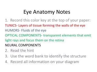
The anatomical position refers to the body position as if • the person were standing upright with the: • Head, eyes, and toes directed anteriorly (forward) • Arms adjacent to the sides with the palms facing anteriorly • Lowerlimbs close together with the feet parallel.
Terms of movementmayalso be considered in pairs of oppositingmovements: Flexionandextensionmovementsgenerallyoccur in sagittalplanesaround a transverseaxis.
@ a frontalplanearound an anteroposterioraxis Abductionmovingawayfromthemedianplaneexceptdigits Adductionmovingtowardsthemedianplane abduction & adduction
The skeletal system may be divided into • 2 functional parts: • The axial skeleton • head (cranium or skull) • neck (hyoid bone and cervical vertebrae) • trunk (ribs, sternum, vertebrae, and sacrum) • The appendicular skeleton • Limbs • including those forming the shoulde & pelvic girdles
HISTOLOGY OF THE BONE sparse cells surrounded by an extracellular network/matrix • 60% the weight of the bone mineral • Rest- water & matrix. • 90% of the matrix proteins collagen1/3 of the bone weight • very strong • forms bone, cartilage, skin, and tendons.
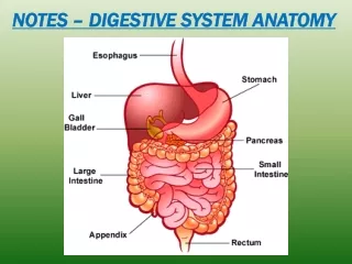
CartilageS and Bones • The skeleton is composed of cartilages and bones. • Cartilage • resilient, semirigid form of connective tissue • forms parts of the skeleton where more flexibility is required. articulating of bones participating in a synovial joint capped with articular cartilage provides smooth, low-friction, gliding surfaces for free movement
Types of cartilage 1. Hyaline most common, matrix w/ moderate amount of collagen fibers articular surfaces of bones 2. Elastic large number of elastic fibers external ear 3. Fibrocartilage limited number of cells & ground substance amidst substantial amount of collagen fibers intervertebral discs
Bones function as • supportive structures for the body • protectors of vital organs • reservoirs of calcium and phosphorus • levers on which muscles act to produce movement • containers for blood-producing cells
TYPES OF BONES • according to their shape gross anatomy • Long bones • tubular humerus in the arm • 3)Flat bones • protectivefunctions • flat bones of the cranium protect the brain 2)Short bones cuboidal tarsus (ankle) carpus (wrist) • 4) Irregular bones • various shapes other than long, short, or flat • bones of the face
5) Sesamoid bones patella or knee cap protect the tendons from excessive wear often change the angle of the tendons as they pass to their attachments.
2 types of bones according to histological features • compact bone & spongy (trabecular) bone • Spongy bone • found @ expanded heads of long bones + fills most irregular bones. • Compact bone • forms outer shell of all bones+shafts in long bones.
skull bones 8 bones of neurocranium
Cranial Fossae Anterior cranial fossa occupiedbytheinferior and anterior parts of the frontal lobes of the brain shallowest cranial fossa Middle cranial fossa butterfly-shaped central part composed of the sellaturcica on the body of the sphenoid large, depressed lateral parts on each side Posterior cranial fossa largest and deepest cranial fossa formed mostly by the occipital bone
Ribs (L. costae) • curved flat bones • form most of the thoracic cage. • 3 types of ribs: • True (vertebrocostal) ribs (1st-7th ribs): • directly to the sternum. • False (vertebrochondral) ribs • (8th, 9th, and usually 10th ribs): • indirect withthesternum • Floating (vertebral, free) ribs • (11th, 12th, and sometimes 10th ribs): • No connectionwiththesternum
STERNUM G. sternon, chest Has threeparts: 1. Manubrium 2. Body 3. Xiphoidprocess
Vertebral column • In an adult typically consists of 33 vertebrae arranged in five regions: 7 cervical, 12 thoracic, 5 lumbar, 5 sacral, and 4 coccygeal. vertebral body vertebral arch seven processes
Clavicle (Tr. Köprücükkemİğİ) • Its medial half articulates with the manubrium of the sternum. • Its lateral half articulates with the scapula. • These curvatures increase the resilience of the clavicle and give it the appearance of an elongated capital S.
Scapula (Tr. Kürekkemiği) • The scapula (shoulder blade) is a triangular flat bone that lies on the posterolateral aspect of the thorax. • The scapula has an articular surface; a glenoid cavity (G. socket) for the articulation with the head of the humerus.
HUMERUS • largest bone in the upper limb • articulates with the scapula at the glenohumeral joint • articulates with the radius and ulna at the elbow joint. • The proximal end of the humerus has a head, surgicaland anatomical necks, and greater and lesser tubercles.
ULNA • stabilizing bone of the forearm • medial and longer of the two forearm bones. Its more massive proximal end is specialized for articulation with the humerus proximally and the head of the radius laterally.
lateral and shorter of the two forearm bones. • Its proximal end includes a short head, neck. RADIUS • Proximally, the head of the radius is concave for articulation with the humerus during flexion and extension of the elbow joint. • The head also articulates with the ulna. • The shaft of the radius, in contrast to that of the ulna, gradually enlarges as it passes distally.
The metacarpus forms the skeleton of the palm of the hand between the carpus and the phalanges. • It is composed of five metacarpal bones (metacarpals). • The proximal bases of the metacarpals articulate with the carpal bones, and the distal heads of the metacarpals articulate with the proximal phalanges and form the knuckles.
The skeleton of the lower limb (inferior appendicular skeleton) may be divided into two functional components: pelvic girdle bones of the free lower limb.
In the mature individual, the pelvic girdle is formed by three bones: Right and left hip bones (coxal bones; pelvic bones): large, irregularly shaped bones, each of which develops from the fusion of three bones, the ilium, ischium, and pubis.
Tibia (Shine bone) on the anteromedial side of the leg, nearly parallel to the fibula • second largest bone • anterior border of the tibia -most prominent border. • tibia & adjacent medial surface subcutaneous throughout their lengths • commonly known as the “shin” • periosteal covering and overlying skin vulnerable to bruising.
Fibula posterolateral to the tibia • slender • tibiofibularsyndesmosis • no function in weight-bearing • serves mainly for muscle attachment. • distal end enlarges prolonged as lateral malleolus • proximal end an enlarged head superior to a small neck.
patella (knee cap) • largest sesamoid bone in the body • embedded in the quadriceps femoris tendon. • jointbetweenthepatellaandfemursharethesamearticularcavity w/ thejointbetweenfemur & tibia • patellar ligament connects the patella to the tibia.
Bones of the foot Tarsus (7 bones) Metatarsus (5 bones) Phalanges (14 phalanges)
according to the tissues that lie between the bones: • Fibrous joints • Syndesmosis type of fibrous joint • 2) Cartilaginous joints • 3) Synovial joints Classification of Joints
Types of synovial joints • according to shape of articulating surfaces- type of movement they permit • Plane joints • uniaxialjoints- glidingorsliding • acromioclavicular joint • 2. Hinge joints • uniaxialjoints- flexion & extension • knee & elbowjoints
Types of synovial joints 3. Saddle joints biaxialjoints- flexion & extension, abduction & adduction carpometacarpal joint at the base of the 1st digit (thumb) 4. Condyloid (ellipsoid type) biaxialjoints- flexion & extension, abduction & adduction metacarpophalangeal joints (knuckle joints) radiocarpal joint (wrist)
Types of synovial joints 5. Ball and socket joints (spheroidaljoints) multiple axes and planes: flexion and extension, abduction and adduction, medial and lateral rotation, and circumduction hip & shoulderjoints
Types of synovial joints 6. Pivot joints uniaxialjoints- rotationaround a centralaxis proximal& distal radioulnar joints
TEMPOROMANDIBULAR JOINT • mandibular fossa &articular tubercle of temporal bone • head of the mandible • articular disc of the TMJ
JOINTS OF THE VERTEBRAL COLUMN • The vertebral column in an adult typically consists of 33 vertebrae arranged in five regions: 7 cervical, 12 thoracic, 5 lumbar, 5 sacral, and 4 coccygeal. • Joints of the vertebral bodiessymphyses (secondary cartilaginous joints) • Joints of the vertebral arches(facetjoints) • Craniovertebral(atlanto-axial and atlanto-occipital) joints • Costovertebraljoints • Sacroiliac joints
JOINTS OF THE UPPER LIMB • Sternoclavicularjoint (SC) • sternalend of the clavicle articulates with manubrium & 1st costal cartilage • Theonlyarticulationbetweenupperlimb& axialskeleton. • During full elevation of the limb, clavicle is raised to 60° angle.
Glenohumeral(shoulder) joint permits a wide range of movement; mobility makes the joint relatively unstable. Humeralhead articulates w/ glenoidcavity of the scapula deepened slightly but effectively by the ring-like, fibrocartilaginousglenoid labrum (L., lip).
Glenohumeral(shoulder) joint more freedom of movement than any other joint in the body • results from the laxity of its joint capsule & large size of the humeral head compared with the small size of the glenoid cavity. • movements around three axes • flexion-extension, abduction-adduction, rotation (medial and lateral) of the humerus, circumduction
ElbowJoint • located inferior to the epicondyles of the humerus • humeroulnar&humeroradialarticulations
Proximal(superior) radio-ulnar joint allows movement of the head of the radius on the ulna Radialhead is held in position by the anular ligament of the radius. Distal(inferior) radio-ulnar joint The radius moves around the relatively fixed distal end of the ulna.
Wrist(radiocarpal) joint ulna does not participate in the wrist joint. Distalend of the radius & articular disc of the distal radio-ulnar joint articulate with proximal row of carpal bones, except for the pisiform. Flexion Extension Abduction Adduction radialdeviation-ulnardeviation Circumduction
JOINTS OF THE LOWER LIMB • articulations of the pelvic girdle • lumbosacral joints, sacroiliac joints, and pubic symphysis • hip joints • knee joints • tibiofibular joints • ankle joints • foot joints
JOINTS OF THE PELVIS Pubicsymphysis interpubicdisc& surroundingligaments unitethebodies of thepubicbones in themedianplane. Lumbosacraljoints L5 and S1 vertebrae articulate Sacrococcygeal joint
Types of Muscles • based on distinct characteristics • Functional • voluntary vs. involuntary • Histological • striated vs. smooth or unstriated • Anatomical (location) • @ body wall (soma) and limbs • @ hollow organs (viscera) or blood vessels