Download
1 / 1
10 likes | 128 Vues
Pregnancy Evaluation of Ovine Clone Obtained by Somatic Cell Nuclear Transfer A.K. Tarouco 1 , A.S. Traldi 2 , M.S. Miranda 1 , D.R. Catto 3 , R.O.C. Silva 3 , A.C. Marchesine 4 , M. R. Watanabe 5 , F.V. Meirelles 1.

E N D
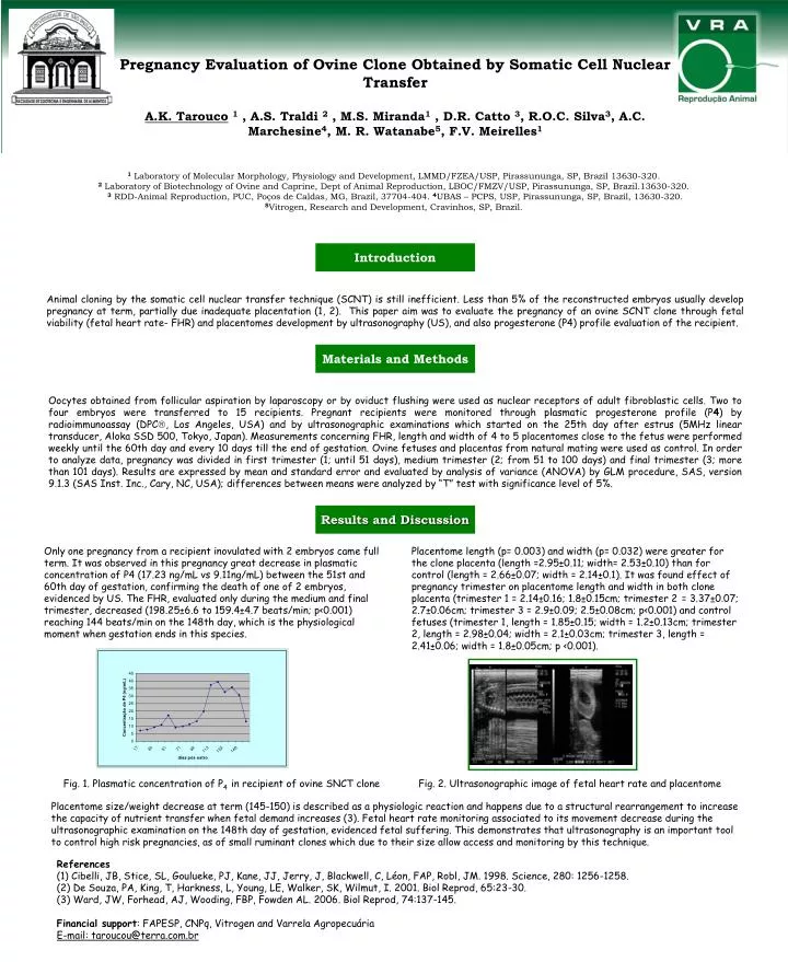
Pregnancy Evaluation of Ovine Clone Obtained by Somatic Cell Nuclear Transfer A.K. Tarouco1 , A.S. Traldi 2 , M.S. Miranda1 , D.R. Catto 3, R.O.C. Silva3, A.C. Marchesine4, M. R. Watanabe5, F.V. Meirelles1 1 Laboratory of Molecular Morphology, Physiology and Development, LMMD/FZEA/USP, Pirassununga, SP, Brazil 13630-320. 2 Laboratory of Biotechnology of Ovine and Caprine, Dept of Animal Reproduction, LBOC/FMZV/USP, Pirassununga, SP, Brazil.13630-320. 3 RDD-Animal Reproduction, PUC, Poços de Caldas, MG, Brazil, 37704-404. 4UBAS – PCPS, USP, Pirassununga, SP, Brazil, 13630-320. 5Vitrogen, Research and Development, Cravinhos, SP, Brazil. Introduction Animal cloning by the somatic cell nuclear transfer technique (SCNT) is still inefficient. Less than 5% of the reconstructed embryos usually develop pregnancy at term, partially due inadequate placentation (1, 2). This paper aim was to evaluate the pregnancy of an ovine SCNT clone through fetal viability (fetal heart rate- FHR) and placentomes development by ultrasonography (US), and also progesterone (P4) profile evaluation of the recipient. Materials and Methods Oocytes obtained from follicular aspiration by laparoscopy or by oviduct flushing were used as nuclear receptors of adult fibroblastic cells. Two to four embryos were transferred to 15 recipients. Pregnant recipients were monitored through plasmatic progesterone profile (P4) by radioimmunoassay (DPC, Los Angeles, USA) and by ultrasonographic examinations which started on the 25th day after estrus (5MHz linear transducer, Aloka SSD 500, Tokyo, Japan). Measurements concerning FHR, length and width of 4 to 5 placentomes close to the fetus were performed weekly until the 60th day and every 10 days till the end of gestation. Ovine fetuses and placentas from natural mating were used as control. In order to analyze data, pregnancy was divided in first trimester (1; until 51 days), medium trimester (2; from 51 to 100 days) and final trimester (3; more than 101 days). Results are expressed by mean and standard error and evaluated by analysis of variance (ANOVA) by GLM procedure, SAS, version 9.1.3 (SAS Inst. Inc., Cary, NC, USA); differences between means were analyzed by “T” test with significance level of 5%. Results and Discussion Only one pregnancy from a recipient inovulated with 2 embryos came full term. It was observed in this pregnancy great decrease in plasmatic concentration of P4 (17.23 ng/mL vs 9.11ng/mL) between the 51st and 60th day of gestation, confirming the death of one of 2 embryos, evidenced by US. The FHR, evaluated only during the medium and final trimester, decreased (198.25±6.6 to 159.4±4.7 beats/min; p<0.001) reaching 144 beats/min on the 148th day, which is the physiological moment when gestation ends in this species. Placentome length (p= 0.003) and width (p= 0.032) were greater for the clone placenta (length =2.95±0.11; width= 2.53±0.10) than for control (length = 2.66±0.07; width = 2.14±0.1). It was found effect of pregnancy trimester on placentome length and width in both clone placenta (trimester 1 = 2.14±0.16; 1.8±0.15cm; trimester 2= 3.37±0.07; 2.7±0.06cm; trimester 3 = 2.9±0.09; 2.5±0.08cm; p<0.001) and control fetuses (trimester 1, length = 1.85±0.15; width = 1.2±0.13cm; trimester 2, length = 2.98±0.04; width = 2.1±0.03cm; trimester 3, length = 2.41±0.06; width = 1.8±0.05cm; p <0.001). Fig. 1. Plasmatic concentration of P4 in recipient of ovine SNCT clone Fig. 2. Ultrasonographic image of fetal heart rate and placentome Placentome size/weight decrease at term (145-150) is described as a physiologic reaction and happens due to a structural rearrangement to increase the capacity of nutrient transfer when fetal demand increases (3). Fetal heart rate monitoring associated to its movement decrease during the ultrasonographic examination on the 148th day of gestation, evidenced fetal suffering. This demonstrates that ultrasonography is an important tool to control high risk pregnancies, as of small ruminant clones which due to their size allow access and monitoring by this technique. References (1) Cibelli, JB, Stice, SL, Goulueke, PJ, Kane, JJ, Jerry, J, Blackwell, C, Léon, FAP, Robl, JM. 1998. Science, 280: 1256-1258. (2) De Souza, PA, King, T, Harkness, L, Young, LE, Walker, SK, Wilmut, I. 2001. Biol Reprod, 65:23-30. (3) Ward, JW, Forhead, AJ, Wooding, FBP, Fowden AL. 2006. Biol Reprod, 74:137-145. Financial support: FAPESP, CNPq, Vitrogen and Varrela Agropecuária E-mail: taroucou@terra.com.br