Download
1 / 50
530 likes | 851 Vues
PATHOPHYSIOLOGY OF SELECTED AUTOIMMUNE DISEASES. Lecture from pathological physiology November 11, 2004. Autoimmunity and autoimmune diseases. Autoimmunity = is an acquired immune reactivity to „self“ components (antigens) Autoimmune diseases

E N D
PATHOPHYSIOLOGY OF SELECTED AUTOIMMUNE DISEASES Lecture from pathological physiology November 11, 2004
Autoimmunity and autoimmune diseases Autoimmunity = is an acquired immune reactivity to „self“ components (antigens) Autoimmune diseases - occur when autoimmune responses lead to tissue damage (functional and morphologic changes)
Autoimmunity • Original conception - autoimmunity = unfavourable phenomena „horror autotoxicus“ • Normal process – eliminate self antigens (aggrieved or inappropriate) autoreactiveT-cells autoantibodies
Self tolerance • lack of immune responsiveness to an individual´s own tissue antigens 3 proposed mechanisms for developing self-tolerance: a) Clonal deletion b) Clonal anergy c) Peripheral suppression central tolerance x peripheral tolerance
Clonal deletion • loss of clones of T and B cells during maturation via apoptosis • more operative for B cells than T cells) Positive and negative selection of T-cells in thymus
Clonal anergy • irreversible loss of function of lymphocytes induced by long-term enconter of antigens. T-cell activation requires to signals. Absence of second signal (from antigen presenting cells) leads to anergy. Nečas et al., 2000
Peripheral suppression by T-cells Suppressor T cells (possibly via IL-10) inactivate T helper and B lymphocytes Nečas et al., 2000
Causes for loss of self-tolerance • Genetic predisposition • Imbalance of suppressor-helper T cell function • Apoptosis • Modification of antigen (via drugs, microorganisms) • Molecular mimicry (infectious agents appear similar to self antigens) • Polyclonal lymphocyte activation (endotoxins cause such activation independent of specific antigens) • Emergence of sequestered antigen
GENETIC PREDISPOSITIONAssociation of selected autoimmune diseases with HLA Diseases HLA Risk* Ankylosing spondylitis B27 90 Reiter´s syndrom B27 36 SLE DR3 15 Myasthenia gravis DR3 2.5 IDDM DR3/DR4 25 Psoriasis vulgaris DR4 14 Multiple sclerosis DR2 5 Rheumatoid arthritis DR4 4 * Based on comparison of the incidence of the autoimmune disease in patients with a given HLA type with the incidence of the autoimmune disease in patients without this HLA type
ROLE OF TH1/TH2 balance • Original response is associated with dominance Th1 or Th2 cytokines • balance Th1 a Th2 autoimmune pathogenesis Th1 is involved in autoimmunity; TH2 has a protective effect. TH1 cells transfer EAE to nonimmunized animals. TH2 protect mice against EAE following immunization with MBP+CFA. e. g- In gravidity (predominance Th2 cytokines) • Th1 autoim. disease RA improve • Th2 autoim. disease SLE grow worse Krejsek et al., 2004
MOLECULAR MIMICRY Role of microbial or viral antigens identical or similar as self cells it leads to reaction against self tissues by same mechanism which removed pathogenes Krejsek et al., 2004
Cross-reacting antibodies play a role in heart damage in rheumatic fever
RELEASE OF SEQUESTERED ANTIGENS As discussed, the induction of self-tolerance in T cells is thought to result from exposure of immature thymocytes to self-antigens and the subsequent clonal deletion of those that are self-reactive. Any tissue antigens that are sequestrated from the circulation, and therefore are not seen by the developing T cells in the thymus, will not induce self-tolerance. Exposure of mature T cells to such normally sequestrated antigens at a later time might result in their activation.
Release of sequestered antigen A few tissue antigens are known to fall into this category. For example: • -MBP following a viral or bacterial infection which affects the brain-blood-barrier; • -sperm following vasectomy • -eye lens proteins following eye damage • -heart muscle Ags following myocardial infarction
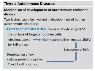
INNAPROPRIATE EXPRESSION OF CLASS II MHC MOLECULES For example: • The pancreatic beta cells of individuals with IDDM express high levels of both class I and class II MHC molecules, whereas healthy beta cells express lower levels of class I and do not express class II at all. • Similarly, thyroid acinar cells from those with Graves´ disease have been shown to express class II MHC molecules on their membranes. This inappropriate expression of class II MHC molecules, which are normally expressed only on antigen-presenting cells, may serve to sensitize TH cells to peptides derived from the beta cells or TC cells or sensitization of TDTH cells against self-antigens.
POLYCLONAL LYMPHOCYTE ACTIVATION • A number of viruses and bacteria can induce nonspecific polyclonal B-cell activation (G- bacteria, CMV, EBV) inducing the proliferation of numerous clones of B cells that express IgM in the absence of TH-ly • If B cells reactive to self-antigens are activated by this mechanism, auto-antibodies can appear.
IgG and IgM- mediated type II hypersensitivity Antibody (IgM or IgG) directed mainly to cellular antigens (e. g. on ERY) or surface autoantigens can cause damage through • opsonization • lysis by complement • antibody dependent cellular cytotoxicity Also called cytotoxic hypersensitivity. Nečas et al., 2000
Immune-complex mediated type III hypersensitivity Immune complexes can form to serum products as well as microbial and self antigens, either in local sites or systemically, leading to phagocytic and complement mediated damage. Tissue damage is caused mainly by complement activation and release of lytic enzymes from neutrophils • local damage (Arthus reaction) • systemic complexes deposition in blood vessels (vasculitis)
Delayed-type (type IV) hypersensitivity This occurs from 24 h after contact with an antigen and is mediated by T cells together with dendritic cells, macrophages and cytokines. Such responses often lead to the production of granulomas as an expression of chronic stimulation of T cells and macrophages, where there is persistance of Ag which the immune system is unable to remove
Type V hypersensitivity It is an example of hypesensitive reaction against cell receptors: • AutoAbs againsthe acetylcholinereceptor found on motor end plates of muscle. Prevent binding of Ach, and induce C-mediated degradation of the receptors (e. g. myasthenia gravis) • AutoAbs against the TSH-receptor bind to the receptor and act as TSH agonists, inducing production of the thyroid hormones (e. g. Graves´ disease)
Autoimmune diseases Autoimmune diseases form a spectrum ranging from organ-specific conditions in which one organ only is affected to systemic diseases in which the pathology is diffused throughout the body. The extremes of this spectrum result from quite distinct underlying mechanisms, but there are many conditions in which there are components of both organ-specific and systemic damage.
Acute infection and autoimmune diseases Krejsek et al., 2004
Reumatoid arthritis (RA) - pathogenesis • Common disease (1% of population), in general in women 40-60 years old • It is characterized by persistent inflammation of the synovium leading to varying degrees of joint destruction. • Genetic predisposition (HLA DRB) – molecular mimicry (e. g. EBV) Krejsek et al., 2004
Reumatoid arthritis - pathogenesis Krejsek et al., 2004
RA – mechanisms and clinical symptoms: • The disease characteristically starts in the small joints (although spares the distal interphalangeal joints) and then spreads to involve more proximal joints. • The synovial membrane undergoes infiltration by lymphocytes (lymphoid follicles arise) causing villous hypertrophy. • MHC class II molecules are strongly expressed on B cells and synovial lining cells. It is thought that the autoantigen is presented to T cells at this site and that AutoAbs production results in immune complex formation. These are phagocytosed by macrophages and neutrophils, leading to their activation, formation of reactive oxygen intermediates and release of lysosomal enzymes and thus tissue damage. • Rheumatoid factors are IgM which react with the Fc. Complexes are deposited in joints and lead to type III hypersensitivity
Ankylosing spondylitis • This is also called „seronegative arthritide“ • Especially in young men • The occurence is clearly associated with HLA-B27 • Symptoms: chronic inflammation, fibrosis, and ossification of the articulations of the spine • Extraarticular symptoms: uveitis (iritis), perikarditis, uretritis Folsch et al., 2003
Systemic lupus erythematosus (SLE) • In general in women 20-40 years old, ratio of female/male 9:1 • Symptoms: fever, weakness, arthritis, skin rashes, pleurisy, kidney dysfunction...) • autoAbs to DNA, histones, RBC, platelets, leukocytes, clotting factors (ANA- AntiNuclear Antibodies) • Deposits of Ag-Ab complexes (type III hypersensitivity) and complement activation cause damage of blood vessel walls, occlusions of small blood vessels, tissue damage Krejsek et al., 2004
SLE – a mouse strain called MRL/lpr/lpr • These mice are homozygous for a gene lrp, which has been identified as a defective fas gene (the fas-gene product is a cell-surface protein belonging to the TNF family receptors) • When the normal Fas protein interacts with its ligand, it transduces a signal that leads to apoptotic death of the Fas-bearing cells. norma: Fas protein + ligand • In the absence of Fas, mature peripheral T cells do not die, and these activated cells continue to proliferate and produce cytokines that result in grossly enlarged lymph nodes and spleen
Scleroderma • Scleroderma is a state of dysregulated connective tissue deposition. It is characterised by expansion of dysregulated fibroblast clones which behave autonomously and overexpress genes encoding elements of the extracellular matrix, particularly type I collagen • 2 forms: - limited (skin) - CREST syndrome (Calcinosis, Raynaud´s phenomenon, Esophageal dysmotility, Sclerodactyly, Teleangiectasia) - diffuse (systemic) – pulmonary fibrosis, renal involvement • AutoAbs to topoisomerase I (Scl-70), to RNA polymerase III AutoAbs to centromera (in CREST syndrome)
Poly- and dermatomyositis • The inflammatory myopathies, PM and DM are important and serious causes of muscle weakness * Muscle weakness– with fiber degeneration, regeneration and widespread infiltration of mononuclear cells * Skin symptoms – inflammatory dermatitis on extensor srfaces of the knuckles (Gottron´s papules) and a violaceous discoloration of the eyelids (heliotrope) • Can be as a paraneoplastic syndrome
Myasthenia gravis • It is the prototype autoimmune disease mediated by blocking auto-antibodies • A patient withthis disease produces autoAbs to the Ach receptors on the motor end-plates of muscles • Binding of these AutoAbs to the receptors blocks the normal binding of Ach and also induces complement-mediated degradation of the receptors, resulting in progressive weakening of the skeletal muscles
Frequently associated with hyperplasia of thymus or thymoma • Association with other autoimmune diseases • Several types (e. g. Eaton-Lambert´s syndrome) Symptoms: • The early signs of this disease include drooping eyelids and inability to retract the corners of the mouth, which gives the appearance of snarling • Progressive weakening of the skeletal muscles Th: antagonists of cholinesterase, thymectomy, immunosuppressive drugs
Multiple sclerosis • An autoimmune disease that affects the CNS • Prevalence 1:1000, most people between the ages of 20 and 40 genetic background x environmental influences (MS is more common in the Northern hemisphere) • Autoantigens: MBP, MAG, MOG, PLP… • Individuals produce autoreactive T cells that participate in the formation of inflammatory lesion along the myelin sheath of nerve fibers T cells cause inflammatory lesions, destroying the myelin. It leads to numerous neurologic dysfunctions
Autoimmune anemias Pernicious: • autoAbs to the membrane-bound intrinsic factor which facilitates the uptake of vit. B12 from the small intestine (owing to autoimmune gastritis type A) Autoimmune hemolytic: • autoAbs to RBC antigens induce complement-mediated lysis of erythrocytes or AutoAbs mediated fagocytosis of ERY Drugs-induced hemolytic anemias: • penicillin, methyldopa e.g. interact with erythrocytes
Goodpasture syndrome • Rare, mostly in young men • Frequently after viral infection • AutoAbs anti-basement membrane Ags for kidney glomeruli and alveoli of lungs (collagen type IV) • Induce complement activation and cellular damage Clinical symptoms: - anemias - progressive kidney damage and pulmonary gemmorrhage - Death within months Therapy: corticosteroids, plasmapheresis
Graves´ disease • Known as Basedow´s disease in some parts of the world. • It is the most common condition that produces hyperthyroidism (about 70%) • This disease is more common in women compared to men by about 10 to one
Pathogenesis The production of autoAbs that bind and activate the thyroid cell surface receptor for TSH. These so-called thyroid-stimulating immunoglobulins (TSIs) TSIs cause production and release of thyroid hormone that is autonomous of regulation by TSH
Klinický obraz: • klasicky: goiter, tachycardia, exopthalmus • hypermetabolic syndrome: K (kůže- skin) – warm, sweating, hair is thin L (labor) - fatique, muscle atrophy M (metabolic symptoms) – weight loss, increased appetite N (nervous symptoms) – nervousness, palpitations, O (oběhové – circulatory symptoms) – tachycardia (or atrial fibrilation), palpitations P (protrusion) – symptoms of infiltrative ophthalmopathy exophthalmosin which the eyes protrude owing to inflammatory and cellular involvement of the retro-orbital tissues, including ocular muscles and fat
Hashimoto thyroiditis • Autoimmune disease mostly in middle aged women • Association with HLA type exists (HLA-DR5, DR-3) • pathogenesis: • Infiltration of lymphocytes, macrophages, plasma cells in the thyroid leads to an inflammatory response which causes a goiter • AutoAbs to thyroglobulin and thyroid peroxidase interfere with iodine uptake and lead to a reduced production of thyroid hormones - symptoms: goiter, breakdown of function (euthyreoidism, hyper- , hypo-)
Insulin-dependent diabetes mellitus Krejsek et al., 2004
Insulin-dependent diabetes mellitus • Autoimmune process causes destruction of cells in the pancreas resulting in insufficient insulin production • LADA (latent immune diabetes adults) – run slowly Th: recombinant human insulin continuous monitoring levels of blood glucose and insulin transplantation of pancreas?
Sjögren´s syndrome • It is a relatively common autoimmune disorder characterised by exocrinopathy resulting in the cardinal manifestations of keratoconjunctivitis sicca (90%) and xerostomia (80%) - sicca syndrome • When these manifestations occur in the absence of another clearly defined connective tissue disease, the diagnosis is primary Sjögren´ s syndrome. • Secondary Sjögren´ s syndrome may occur in association with a variety of other autoimmune diseases. • Women are disproportionately affected (90%) • Interaction between genetic (HLA B8, DR3, DR2) and environmental factors
Clinical symptoms Glandular manifestations: • Keratoconjunctivitis sicca (dry eyes, grittiness, burning, photophobia..) • Xerostomia (dry mouth, odynophagia, halitosis, dysgeusia…) Extraglangular manifestations: • Respiratory diseases – interstitial diseases… • Renal diseases – intersticial nephritis, tubular dysfunction • Neurological – peripheral or cranial neuropathy • Arthritis • Cutaneous vasculitis