Download
1 / 20
230 likes | 476 Vues
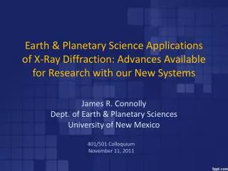
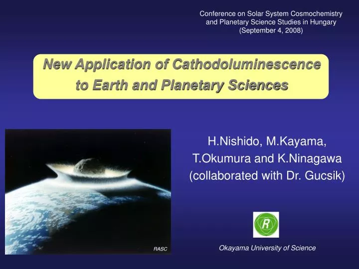
Conference on Solar System Cosmochemistry and Planetary Science Studies in Hungary (September 4, 2008). New Application of Cathodoluminescence to Earth and Planetary Sciences. H.Nishido, M.Kayama, T.Okumura and K.Ninagawa (collaborated with Dr. Gucsik) Okayama University of Science. RASC.

E N D
Conference on Solar System Cosmochemistry and Planetary Science Studies in Hungary (September 4, 2008) New Application of Cathodoluminescence to Earth and Planetary Sciences H.Nishido, M.Kayama, T.Okumura and K.Ninagawa (collaborated with Dr. Gucsik) Okayama University of Science RASC
Different types of luminescence Type of luminescence Excitation sourceApplication to geosciences Thermoluminescence (TL) Thermal energy Dosimeter, Dating Optically Stimulated Luminescence (OSL) Visible light Dosimeter, Dating Photoluminescence (PL) Ultraviolet radiation Mining exploration Cathodoluminescence (CL) Electrons Fabric and domain analysis Roentgenoluminescence (RL) X-rays Dosimeter Ionoluminescence Ions Dosimeter Chemiluminescence Chemical reactions Water circulation analysis Bioluminescence Biochemical reactions Detection of micro biomats Triboluminescence Shear stress Bedrock monitoring sensor
Different types of luminescence Type of luminescence Excitation sourceApplication to geosciences Thermoluminescence (TL) Thermal energy Dosimeter, Dating Optically Stimulated Luminescence (OSL) Visible light Dosimeter, Dating Photoluminescence (PL) Ultraviolet radiation Mining exploration Cathodoluminescence (CL) Electrons Fabric and domain analysis Roentgenoluminescence (RL) X-rays Dosimeter Ionoluminescence Ions Dosimeter Chemiluminescence Chemical reactions Water circulation analysis Bioluminescence Biochemical reactions Detection of micro biomats Triboluminescence Shear stress Bedrock monitoring sensor
Different types of luminescence Type of luminescence Excitation sourceApplication to geosciences Thermoluminescence (TL) Thermal energy Dosimeter, Dating Optically Stimulated Luminescence (OSL) Visible light Dosimeter, Dating Photoluminescence (PL) Ultraviolet radiation Mining exploration Cathodoluminescence (CL) Electrons Fabric and domain analysis Roentgenoluminescence (RL) X-rays Dosimeter Ionoluminescence Ions Dosimeter Chemiluminescence Chemical reactions Water circulation analysis Bioluminescence Biochemical reactions Detection of micro biomats Triboluminescence Shear stress Bedrock monitoring sensor Quartz images by polarizing microscopy (Polmi) and SEM‐CL microscopy (CL), Götze (2000).
Cold-cathode type CL instrument (Luminoscope) ・ Color imaging of CL ・ Comparison with petrological observation Luminoscope ELM-3R (Nuclide Co.) Cooled-CCD system: DS-5Mc(Nikon Co.) Optical system: Video lens (Edmund Co.)
Phase II Phase III CLimageofAntarctic meteorite Phase I E-type chondrite (Y-86004) width: 4.5 mm Courtesy of Prof. Ninagawa
Monochromator Hot-cathode type CL instrument (SEM-CL) ・ CL spectral measurement ・ Comparison with SEM and BSE images, linked to EPMA SEM: JSM-5410LV (JEOL Co.) Monochromator: Mono CL2 (Oxford Instruments Co.) PMT: R2228 (Hamamatsu PhotonicsCo.) Wavelength: 300~800 nm Resolution: 0.5 nm Temperature: -196~400 C
CL characterization of zoned zircon A C D B Zircon from Osayama, Okayama Pref., Japan A: Luminoscope CL image; B: Mono-CL image; C: CL spectral measurement positions; D: CL spectra
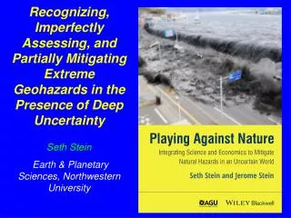
shatter cone Conditions of shock metamorphism (from French, 1998) Barringer impact crater, Arizona
Shocked quatrz Meteor Impact → shock meatmorphism PF (Planar Fractures) PDFs (Planar Deformation Features) Quartz ・Conventional methods: Optical observation,TEM etc ・New methods: CL imaging and spectroscopy, Micro-Raman spectroscopy PF PDFs 100 mm 50 mm PF and PDFs in quartz grains from Ries crater
Geological setting Location map Geological map
Occurrence a: viewing from NE to SW; b: outcrop; c: chert vein
b a c d Polarized microscope images a, b, d: chert; c: sandstone
a b SEM images of HF etched quartz white arrow: "pillaring" texture; black arrow: "array" texture
PDF images SEM-CL image Optical image SEM image BSE image
Raman spectral analysis Micro-Raman spectra of shocked quartz from Mt. Oikeyama
Optical image SEM-CL image 3D Raman mapping 2D Raman mapping Shocked quartz from Mt. Oikeyama
Bouguer anomaly map Impact crater structure
Concluding Remarks ・ CL method provide us a useful information on defect in the lattice and trace elements existed as a impurity, which are so difficult to characterize using any other conventional methods. ・ SEM-CL and micro-Raman spectroscopy enable to characterize crystallochemical properties of micro-size minerals in planetary science. ・ Further CL application can be expected to a new field in geosciences.