Download

1 / 19
280 likes | 745 Vues
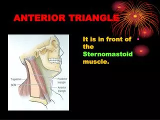
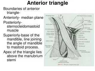
ANTERIOR TRIANGLE OF THE NECK II. Dr. Ahmed Fathalla Ibrahim. CAROTID TRIANGLE. BOUNDARIES: Superiorly: posterior belly of digastric Inferiorly: superior belly of omohyoid Posteriorly: sternomastoid FLOOR: Anteriorly: hyoglossus & thyrohoid muscles

E N D
ANTERIOR TRIANGLE OF THE NECK II Dr. Ahmed Fathalla Ibrahim
CAROTID TRIANGLE BOUNDARIES: • Superiorly: posterior belly of digastric • Inferiorly: superior belly of omohyoid • Posteriorly: sternomastoid FLOOR: • Anteriorly: hyoglossus & thyrohoid muscles • Posteriorly: middle & inferior constrictors of pharynx C
CONTENTS OF CAROTID TRIANGLE CAROTID SHEATH: • A condensation of deep fascia that contains: • Common & internal carotid arteries: (medially) • Internal jugular vein: (laterally) • Vagus nerve: between & a little posterior to arteries & vein
CONTENTS OF CAROTID TRIANGLE • ARTERIES: • Termination of common carotid: at level of disc between C3 & C4 (level of upper border of thyroid cartilage) • Proximal part of external carotid • Proximal part of internal carotid • Five branches of external carotid: superior thyroid, ascending pharyngeal, lingual, facial & occipital
CONTENTS OF CAROTID TRIANGLE • VEINS: • Internal jugular vein • Five tributaries to internal jugular veins: common facial, lingual, superior thyroid, middle thyroid, pharyngeal veins
CONTENTS OF CAROTID TRIANGLE FIVE NERVES: • TWO NERVES RELATED TO CAROTID SHEATH: • Ansa cervicalis (anterior rami of C1,2,3): embedded in the anterior wall of sheath • Sympathetic trunk: embedded in the posterior wall of sheath
CONTENTS OF CAROTID TRIANGLE • THREE NERVES BETWEEN INTERNAL JUGULAR VEIN & INTERNAL CAROTID ARTERY: • Vagus (10th cranial) nerve: descends between internal jugular vein & common carotid artery • Spinal part of accessory (11th cranial) nerve: crosses internal jugular vein, passes deep to sternomastoid to reach posterior triangle • Hypoglossal (12th cranial) nerve: crosses internal & external carotid arteries, passes deep to posterior belly of digastric to reach digastric triangle
CONTENTS OF CAROTID TRIANGLE • DEEP CERVICAL LYMPH NODES: embedded in the anterior wall of sheath, along the internal jugular vein
MUSCULAR TRIANGLE BOUNDARIES: • Anteriorly: midline of neck • Superiorly: superior belly of omohyoid • Inferiorly: sternomastoid M
MUSCULAR TRIANGLE CONTENTS: THE INFRAHYOID MUSCLES • Superficial layer: • Sternohyoid: medially • Omohyoid: laterally • Deep layer: • Thyrohyoid: above • Sternothyroid: below
MUSCULAR TRIANGLE FLOOR: • PRETRACHEAL FASCIA • STRUCTURES IN THE INFRAHYOID PART OF MEDIAN REGION OF FRONT OF NECK: • Thyrohyoid membrane • Thyroid cartilage • Cricothyroid ligament & muscle • Cricoid cartilage • Cricotracheal ligament • Trachea • Structures in front of trachea: jugular arch, inferior thyroid vein, isthmus of thyroid gland
INFRAHYOID MUSCLES(SUPERFICIAL LAYER) Sternohyoid: • Origin: back of manubrium sterni • Insertion: hyoid bone • Action: depression of hyoid bone Omohyoid: • Superior belly:hyoid bone • Inferior belly:upper border of scapula • Insertion: intermediate tendon (deep to sternomastoid) attached to clavicle • Action: depression of hyoid bone
INFRAHYOID MUSCLES(DEEP LAYER) Thyrohyoid: • Origin: oblique line of thryroid cartilage • Insertion: hyoid bone • Action: depression of hyoid bone, elevation of larynx Sternothyroid: • Origin: back of manubrium sterni • Insertion: oblique line of thryroid cartilage • Action: depression of larynx
INFRAHYOID MUSCLES NERVE SUPPLY: • All muscles are supplied by ansa cervicalis (anterior rami of C1,2,3) EXCEPT:thyrohyoid (by anterior ramus of C1)