Download
1 / 32
460 likes | 1.17k Vues
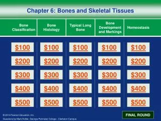
Chapter 6- Part I Bones and Skeletal Tissues. Components of the skeletal system Classification of Bone (bone shapes) Functions of bone Bone structure Microscopic structure of bone and bone cells. What are the parts of the skeletal system?. Bones of the skeleton

E N D
Chapter 6- Part I Bones and Skeletal Tissues • Components of the skeletal system • Classification of Bone (bone shapes) • Functions of bone • Bone structure • Microscopic structure of bone and bone cells
What are the parts of the skeletal system? • Bones of the skeleton • The 206 bones of the body are divided into two categories: • The axial skeleton and the • The appendicular skeleton • Cartilage, ligaments, and other connective tissue that stabilize or connect bones
Growth of Cartilage • Appositional – cells in the perichondrium secrete matrix against the external face of existing cartilage • Interstitial – lacunae-bound chondrocytes inside the cartilage divide and secrete new matrix, expanding the cartilage from within • Calcification of cartilage occurs • During normal bone growth • During old age
Notes on perichondrium perichondrium chondroblast A fibrous membrane covering the external surface of cartilaginous structures The perichondrium nourishes the avascular cartilage, and it also contains mesenchymal cells which can differentiate into chondroblasts. Growth from the perichondrium is appositional growth.
Bones come in many shapes (c) Flat bone (sternum) (a) Long bone (humerus) (b) Irregular bone (vertebra), right lateral view (d) Short bone (talus)
What are some of the functions of bone? • Support • Protection • Movement/leverage • Storage of minerals/fat • Blood cell production
Gross anatomy of bones- Bone markings • Bone surfaces are rarely smooth- they display projections, depressions and openings • WHY? • Projections (or elevations) • Tuberosity, crest, process, crest, spine, condyle etc, etc. • Depressions, grooves, openings in bone • Fossae, sulcus, foramen, grooves, canal, etc. • These are fixed landmarks
Landmarks of the Humerus Parts of the humerus - head, surgical neck, greater tubercle, lesser tubercle, lateral epicondyle, medial epicondyle, trochlea, capitulum, coronoid fossa, olecranon fossa, deltoid tuberosity
trabeculae Compact and Spongy Bone spongy bone • Each bone in the skeleton contains both compactbone and spongy bone. compact bone Compact bone link
Bone structure • Let’s first take a look at the structure of a typical long bone • Then we’ll cover/compare the structure of long bones to short, irregular, and flat bones
Structure of long bones • Diaphysis- tubular shaft, with wall made of compact bone, and a true marrow cavity inside • Epiphysis- each ‘swollen’ end of the bone. Composed largely of spongy bone, with a thin, outer layer of compact bone • The joint surfaces of each epiphysis is covered with hyaline (articular) cartilage • Between the epiphysis and diaphysis (in adult bone)is the epiphyseal line
Periosteum (Periosteum) • Covers outer surface of all bones, except at joint surfaces • Isolates bone from surrounding tissue • Route for nerves and blood supply • Provides an insertion point for tendons/ligaments • Actively participates in bone growth and repair • A double membrane with an outer fibrous layer (dense irregular CT) and an inner cellular layer • Osteoblasts- bone grows thicker
Endosteum (Endosteum) • Covers internal bone surfaces • Including: trabeculae, marrow cavity and canals within compact bone • Contains bone-forming cells, bone destroying cells and stem cells (osteoprogenitor) cells • Active in bone growth, repair and remodeling
Bone Cells • Cells: • Osteoblasts – bone-forming cells • Osteocytes – mature bone cells • Osteoprogenitor cells – stem cells, grandfather cells • Osteoclasts – large cells that resorb or break down bone matrix • More to come
Structure of short, irregular, and flat bones • Consist of thin plates of compact bone on the outside and spongy bone on the inside • Contain bone marrow, but no formal marrow cavity is present • In flat bones, the internal layer of spongy bone is called diploe • Where is the periosteum, endosteum? ..
Where do you find marrow and what is the function? • Red marrow, hematopoietic tissue • Yellow marrow
compact bone The microscopic structure of • Osteon- basic functional unit • Osteocytes arranged around central canal, between concentric lamellae • One small artery, one venule, one nerve, parallel with bone surface • Perforating canal • Perpendicular to long axis of bone, connect blood supply from periosteum through to marrow cavity • Interstitial lamellae • Circumferential lamellae
Spongy bone Compact bone Perforating (Volkmann’s) canal Central (Haversian) canal Endosteum lining bony canals and covering trabeculae Osteon (Haversian system) Circumferential lamellae (a) Perforating (Sharpey’s) fibers Periosteal blood vessel Lamellae Periosteum
A SEM of compact bone Functions of compact bone?
Lamella not in osteons Trabeculae No capillaries within matrix of spongy bone Nutrients reach osteocytes via diffusion through canaliculi that reach surface Trabeculae protect cells of red marrow, yellow marrow Marrow is vascularized The structure of spongy bone Canaliculi opening to surface
Structure of spongy bone Spongy bone (diploë) Compact bone Trabeculae Functions of spongy bone? Link http://depts.washington.edu/bonebio/ASBMRed/structure.html
Composition of Bone (osseous tissue) • Remember that in all connective tissues, there are cells, fibers and ground substance • Cells • Matrix- solid, collagen and Ca2+ deposits • Osteoid makes up 1/3 of bone tissue = collagen fibers, proteoglycans and glycoproteins • Calcium phosphates account for 2/3 the weight of bone • Form hydroxyapatite crystals • Crystals lie in, around, and on the collagen fibers. Fibers form framework for crystals &developing osteocytes
Cells of the Bone- Four major types (a) Osteogenic cell (b) Osteoblast (c) Osteocyte (d) Osteoclast Stem cell Matrix-synthesizing cell responsible for bone growth Mature bone cell that maintains the bone matrix Bone-resorbing cell
Lineage of bone cells Who is missing?
Osteogenic (progenitor) cells • Stem cells of the bone • Produce daughter cells that differentiate into osteoblasts • Cigarette smoke? • Found in endosteum and periosteum
Osteoblasts osteoid • Osteoblasts line all surfaces of bone • Osteoblasts are the builders! • Produce new osteoid • Differentiate into osteocytes • Osteoblasts have receptors for estrogens • Effects of estrogen on these cells?
Osteocytes • Mature bone cells • One osteocyte sits in a lacunae • Cytoplasmic cellular extensions reside in canaliculi • Gap junctions connect neighboring cells • Allow for exchange of nutrients/ions/waste, etc. • Functions- maintain protein/mineral content of matrix, and bone repair
Osteoclasts • Remove/resorb and recycle bone matrix • Found largely in endosteum • Giant cells, 50 or more nuclei • Cousins of macrophage! Do not arise from osteogenic cells • Release acids and digestive enzymes onto matrix • Important in bone remodeling and regulation of blood Ca2+ • Activity inhibited by estrogen
Homeostasis- A balance exists between osteoblast and osteoclast activity LINK • If osteoblasts are working faster than osteoclasts, what happens to bone? • If osteoclasts are working faster, what happens to bone? Breakdown > building? Blood calcium?