Download
1 / 30
300 likes | 535 Vues
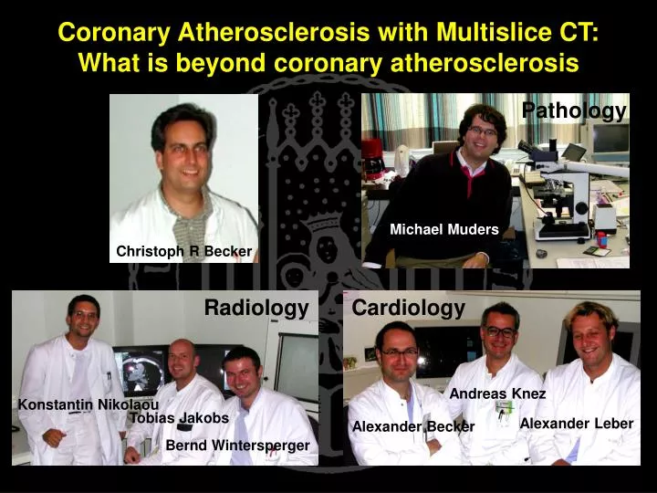
Pathology. Michael Muders. Cardiology. Andreas Knez. Alexander Leber. Alexander Becker. Coronary Atherosclerosis with Multislice CT: What is beyond coronary atherosclerosis. Christoph R Becker. Radiology. Konstantin Nikolaou. Tobias Jakobs. Bernd Wintersperger.

E N D
Pathology Michael Muders Cardiology Andreas Knez Alexander Leber Alexander Becker Coronary Atherosclerosis with Multislice CT:What is beyond coronary atherosclerosis Christoph R Becker Radiology Konstantin Nikolaou Tobias Jakobs Bernd Wintersperger
CTA Inclusion Criteria • Asymptomatic patients • CV risk factors • Positive calcium scan • Symptomatic patients • No CAD history • Atypical chest pain • Inconsistent stress test • 100 mg CaHA
82 bpm 65 bpm Patient Preparation • -blocker • R/o Contra indications • Informed consent • Metoprolol • 50 - 100 mg orally • 30 - 90 min prior • HR 50 - 60 bpm
Coronary CTA Parameters • Testbolus 20 ml @ 4 ml/s • 120 ml (300 mg iodine) @ 3 ml/s + NaCl 60 ml @ 3 ml/s • 500 ms gantry rotation • 120 kV, 300 mA • 4 x 1 mm collimation • 3 mm/s table feed • 40 s breath hold
250 ms 250 ms 250 ms Pitch <0,4 100% 20% mAs ECG Tube Current Modulation
Left Coronary Artery (RAO) Coronary Angiography MDCT & VRT
Right Coronary Artery (LAO) LAO 60 Coronary Angiography MDCT & VRT
Detection of Coronary Stenoses MDCT Coronary Angiography
CTA Limitations • Artifacts • Cardiac motion • Breathing • Blooming • Poor opacification • Small vessel
Solutions • Rot. time & blocker • Cardiac motion artifacts • Scan times • Breathing artifacts • CM utilization • Slice thickness • Small vessels • Blooming artifact
16 Detector Row CT Angiography • 200 ms • 9 Lp/cm • 0.8 mm • 20 s breath hold • 60 ml CM
Coronary Plaque Imaging MDCT Coronary Angiography
Atheroma Rupture/Erosion Healing Hemorrhage Thrombus Calcified Nodule Fibrocalcified Plaque Wall changes Stenoses Occlusion Coronary Atherosclerosis Intimal Thickening
50 HU 50HU Atheroma • 38 YOM • Non specific complain • Risk Factors • Cholesterin • Smoker • No calcium
62 YOM Suspicion of CAD 12 mg CaHA Calcified Nodule
Fibrocalcified Plaque 100 HU
20 HU Thrombus • 42 YOM • Epigastric chest pain • Risk Factors • Hypertension • Smoker • No calcium
Myocardial Infarction Scar anterior LAD posterior RCA lateral LCx
CT Plaque Density p = 0.018 89 ± 31 HU 50 ± 12 HU Lipid Fibrose Lipid Fibrosis
Plaque DistributionLeber 2001 Circulation Myocardial Infarction n = 12 122 Plaque Stable Angina n = 12 135 Plaque
Summary • Detection of stenoses • Calcium • Small vessels • Characterization of plaques • Identify atheromas • Follow up under therapy • Acute coronary event • Intracoronary thrombus • Myocardial infarction