Download
1 / 63
950 likes | 1.83k Vues
Autonomic nervous system. Dilate. Muse lecture #17 Ch 16. Autonomic Nervous System (ANS). The ANS consists of motor neurons that: Innervate smooth and cardiac muscle and glands Make adjustments to ensure optimal support for body activities Operate via subconscious control.

E N D
Autonomic nervous system Dilate Muse lecture #17 Ch 16
Autonomic Nervous System (ANS) • The ANS consists of motor neurons that: • Innervate smooth and cardiac muscle and glands • Make adjustments to ensure optimal support for body activities • Operate via subconscious control
Autonomic Nervous System (ANS) • Other names • Involuntary nervous system • General visceral motor system
Central nervous system (CNS) Peripheral nervous system (PNS) Sensory (afferent) division Motor (efferent) division Somatic nervoussystem Autonomic nervous system (ANS) Sympathetic division Parasympathetic division
Somatic and Autonomic Nervous Systems • The two systems differ in • Effectors • Efferent pathways (and their neurotransmitters) • Target organ responses to neurotransmitters
Effectors • Somatic nervous system • Skeletal muscles • ANS • Cardiac muscle • Smooth muscle • Glands
Efferent Pathways • Somatic nervous system • A, thick, heavily myelinated somatic motor fiber makes up each pathway from the CNS to the muscle • ANS pathway is a two-neuron chain • Preganglionic neuron (in CNS) has a thin, lightly myelinated preganglionic axon • Ganglionic neuron in autonomic ganglion has an unmyelinated postganglionic axon that extends to the effector organ
Neurotransmitter Effects • Somatic nervous system • All somatic motor neurons release acetylcholine (ACh) • Effects are always stimulatory • ANS • Preganglionic fibers release ACh • Postganglionic fibers release norepinephrine or ACh at effectors • Effect is either stimulatory or inhibitory, depending on type of receptors
Neuro- transmitter at effector Cell bodies in central nervous system Effector organs Peripheral nervous system Effect Single neuron from CNS to effector organs ACh + SOMATIC NERVOUS SYSTEM Stimulatory Heavily myelinated axon Skeletal muscle Two-neuron chain from CNS to effector organs NE ACh Unmyelinated postganglionic axon Ganglion SYMPATHETIC Lightly myelinated preganglionic axons + Epinephrine and norepinephrine ACh Stimulatory or inhibitory, depending on neuro- transmitter and receptors on effector organs AUTONOMIC NERVOUS SYSTEM Adrenal medulla Blood vessel ACh ACh Smooth muscle (e.g., in gut), glands, cardiac muscle PARASYMPATHETIC Lightly myelinated preganglionic axon Unmyelinated postganglionic axon Ganglion Acetylcholine (ACh) Norepinephrine (NE) Figure 14.2
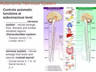
Autonomic Nervous System Figure 16-2b The Organization of the Autonomic Nervous Systems.
Divisions of the ANS • Sympathetic division • Parasympathetic division • Dual innervation • Almost all visceral organs are served by both divisions, but they cause opposite effects
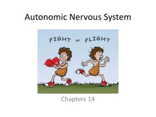
Role of the Parasympathetic Division The brakes • Promotes maintenance activities and conserves body energy • Its activity is illustrated in a person who relaxes, reading, after a meal • Blood pressure, heart rate, and respiratory rates are low • Gastrointestinal tract activity is high • Pupils are constricted and lenses are accommodated for close vision
Role of the Sympathetic Division The gas • Mobilizes the body during activity; is the “fight-or-flight” system • Promotes adjustments during exercise, or when threatened • Blood flow is shunted to skeletal muscles and heart • Bronchioles dilate • Liver releases glucose
Parasympathetic Sympathetic Eye Eye Brain stem Salivary glands Skin* Cranial Salivary glands Sympathetic ganglia Heart Cervical Lungs Lungs T1 Heart Stomach Thoracic Stomach Pancreas Liver and gall- bladder Pancreas L1 Adrenal gland Liver and gall- bladder Lumbar Bladder Bladder Genitals Genitals Sacral Figure 14.3
Eye Ciliary ganglion CN III Lacrimal gland CN VII Pterygopalatine ganglion Pterygopalatine ganglion Nasal mucosa CN IX CN X Submandibular ganglion Submandibular and sublingual glands Otic ganglion Parotid gland Heart Cardiac and pulmonary plexuses Lung Liver and gallbladder Celiac plexus Stomach Pancreas S2 Large intestine S4 Pelvic splanchnic nerves Small intestine Rectum Inferior hypogastric plexus Urinary bladder and ureters Preganglionic Genitalia (penis, clitoris, and vagina) Postganglionic Cranial nerve
Sympathetic (Thoracolumbar) Division • Preganglionic neurons are in spinal cord segments T1 – L2 • Sympathetic neurons produce the lateral horns of the spinal cord • Preganglionic fibers pass through the white rami communicantes and enter sympathetic trunk (paravertebral) ganglia
Eye Lacrimal gland Nasal mucosa Pons Sympathetic trunk (chain) ganglia Blood vessels; skin (arrector pili muscles and sweat glands) Superior cervical ganglion Salivary glands Middle cervical ganglion Heart Inferior cervical ganglion Cardiac and pulmonary plexuses Lung T1 Greater splanchnic nerve Lesser splanchnic nerve Liver and gallbladder Celiac ganglion L2 Stomach Superior mesenteric ganglion White rami communicantes Spleen Adrenal medulla Kidney Sacral splanchnic nerves Lumbar splanchnic nerves Small intestine Inferior mesenteric ganglion Large intestine Rectum Preganglionic Postganglionic Genitalia (uterus, vagina, and penis) and urinary bladder
Sympathetic Trunks and Pathways • There are 23 paravertebral ganglia in the sympathetic trunk (chain) • 3 cervical • 11 thoracic • 4 lumbar • 4 sacral • 1 coccygeal
Spinal cord Dorsal root Ventral root Rib Sympathetic trunk ganglion Sympathetic trunk Ventral ramus of spinal nerve Gray ramus communicans White ramus communicans Thoracic splanchnic nerves (a) Location of the sympathetic trunk
Sympathetic Trunks and Pathways • Upon entering a sympathetic trunk ganglion a preganglionic fiber may do one of the following: • Synapse with a ganglionic neuron within the same ganglion • Ascend or descend the sympathetic trunk to synapse in another trunk ganglion • Pass through the trunk ganglion and emerge without synapsing
Lateral horn (visceral motor zone) Dorsal root Dorsal root ganglion Dorsal ramus of spinal nerve Ventral ramus of spinal nerve Gray ramus communicans Skin (arrector pili muscles and sweat glands) Ventral root White ramus communicans Sympathetic trunk ganglion Sympathetic trunk To effector 1 Synapse at the same level Blood vessels (b) Three pathways of sympathetic innervation
Skin (arrector pili muscles and sweat glands) To effector Blood vessels 2 Synapse at a higher or lower level (b) Three pathways of sympathetic innervation
Splanchnic nerve Collateral ganglion (such as the celiac) Target organ in abdomen (e.g., intestine) 3 Synapse in a distant collateral ganglion anterior to the vertebral column (b) Three pathways of sympathetic innervation
Pathways with Synapses in Chain Ganglia • Postganglionic axons enter the ventral rami via the gray rami communicantes • These fibers innervate • Sweat glands • Arrector pili muscles • Vascular smooth muscle
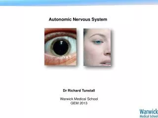
Pathways to the Head • Fibers emerge from T1 – T4 and synapse in the superior cervical ganglion • These fibers • Innervate skin and blood vessels of the head • Stimulate dilator muscles of the iris • Inhibit nasal and salivary glands
Pathways to the Thorax • Preganglionic fibers emerge from T1 – T6 and synapse in the cervical trunk ganglia • Postganglionic fibers emerge from the middle and inferior cervical ganglia and enter nerves C4 – C8 • These fibers innervate: • Heart via the cardiac plexus • Thyroid gland and the skin • Lungs and esophagus
Pathways with Synapses in Collateral Ganglia • Most fibers from T5 – L2 synapse in collateral ganglia • They form thoracic, lumbar, and sacral splanchnic nerves • Their ganglia include the celiac and the superior and inferior mesenteric
Pathways to the Abdomen • Preganglionic fibers from T5 – L2 travel through the thoracic splanchnic nerves • Synapses occur in the celiac and superior mesenteric ganglia • Postganglionic fibers serve the stomach, intestines, liver, spleen, and kidneys
Pathways to the Pelvis • Preganglionic fibers from T10 – L2 travel via the lumbar and sacral splanchnic nerves • Synapses occur in the inferior mesenteric and hypogastric ganglia • Postganglionic fibers serve the distal half of the large intestine, the urinary bladder, and the reproductive organs
Pathways with Synapses in the Adrenal Medulla • Some preganglionic fibers pass directly to the adrenal medulla without synapsing • Upon stimulation, medullary cells secrete norepinephrine and epinephrine into the blood
Visceral Reflexes • Visceral reflex arcs have the same components as somatic reflexes • Main difference: visceral reflex arc has two neurons in the motor pathway • Visceral pain afferents travel along the same pathways as somatic pain fibers, contributing to the phenomenon of referred pain
Stimulus Dorsal root ganglion Sensory receptor in viscera 1 Spinal cord Visceral sensory neuron 2 Integration center • May be preganglionic neuron (as shown) • May be a dorsal horn interneuron • May be within walls of gastrointestinal tract 3 Autonomic ganglion Efferent pathway (two-neuron chain) • Preganglionic neuron • Ganglionic neuron 4 Visceral effector 5 Response
Referred Pain • Visceral pain afferents travel along the same pathway as somatic pain fibers • Pain stimuli arising in the viscera are perceived as somatic in origin
Heart Lungs and diaphragm Liver Heart Gallbladder Liver Appendix Stomach Pancreas Small intestine Ovaries Colon Kidneys Urinary bladder Ureters
Dual Innervation Figure 16–9 Summary: The Anatomical Differences between the Sympathetic and Parasympathetic Divisions.
Neurotransmitters • Cholinergic fibers release the neurotransmitter ACh • All ANS preganglionic axons • All parasympathetic postganglionic axons • Adrenergic fibers release the neurotransmitter NE • Most sympathetic postganglionic axons • Exceptions: sympathetic postganglionic fibers secrete ACh at sweat glands and some blood vessels in skeletal muscles
Two-neuron chain from CNS to effector organs NE ACh Unmyelinated postganglionic axon Ganglion SYMPATHETIC Lightly myelinated preganglionic axons + Epinephrine and norepinephrine ACh Stimulatory or inhibitory, depending on neuro- transmitter and receptors on effector organs AUTONOMIC NERVOUS SYSTEM Adrenal medulla Blood vessel ACh ACh Smooth muscle (e.g., in gut), glands, cardiac muscle PARASYMPATHETIC Lightly myelinated preganglionic axon Unmyelinated postganglionic axon Ganglion Acetylcholine (ACh) Norepinephrine (NE) Figure 14.2
Receptors for Neurotransmitters • Cholinergic receptors for ACh • Adrenergic receptors for NE
Cholinergic Receptors • Two types of receptors bind ACh • Nicotinic • Muscarinic • Named after drugs that bind to them and mimic ACh effects
Nicotinic Receptors • Found on • Motor end plates of skeletal muscle cells (Chapter 9) • All ganglionic neurons (sympathetic and parasympathetic) • Hormone-producing cells of the adrenal medulla • Effect of ACh at nicotinic receptors is always stimulatory
Muscarinic Receptors • Found on • All effector cells stimulated by postganglionic cholinergic fibers • The effect of ACh at muscarinic receptors • Can be either inhibitory or excitatory • Depends on the receptor type of the target organ
Adrenergic Receptors • Two types • Alpha () (subtypes 1, 2) • Beta () (subtypes 1, 2 , 3) • Effects of NE depend on which subclass of receptor predominates on the target organ Beta blockers sometimes given to heart patients
Effects of Drugs • Atropine • Anticholinergic; blocks muscarinic receptors • Used to prevent salivation during surgery, and to dilate the pupils for examination • Neostigmine • Inhibits acetylcholinesterase • Used to treat myasthenia gravis
Effects of Drugs • Over-the-counter drugs for colds, allergies, and nasal congestion • Stimulate -adrenergic receptors • Beta-blockers • Drugs that attach to 2 receptors to dilate lung bronchioles in asthmatics; other uses
Interactions of the Autonomic Divisions • Most visceral organs have dual innervation • Dynamic antagonism allows for precise control of visceral activity • Sympathetic division increases heart and respiratory rates, and inhibits digestion and elimination • Parasympathetic division decreases heart and respiratory rates, and allows for digestion and the discarding of wastes