Download
1 / 41
440 likes | 548 Vues
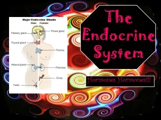
AGING OF THE ENDOCRINE SYSTEM. IEVA B. AKBAR. INTRODUCTION. The aging process can alter neuroendocrine function at multiple levels (i.e. through its effects on biogenic aminergic and peptidergic neurons, anterior pituitary cells and end organs). NEUROTRANSMITTER REGULATION.

E N D
AGING OF THE ENDOCRINE SYSTEM IEVA B. AKBAR
INTRODUCTION The aging process can alter neuroendocrine function at multiple levels (i.e. through its effects on biogenic aminergic and peptidergic neurons, anterior pituitary cells and end organs)
NEUROTRANSMITTER REGULATION There is abundant evidence that biogenic aminergic and peptidergic neurons strongly influence the secretion of hypothalamic hormones. Those most studied are the “classic” neurotransmitter dopamine, norepinephrine, epinephrine and serotonine and the opioids. Other nioamines and peptides have been studied less extensively, and the data are somewhat confounding and inconclusive
DOPAMINE Although histochemical studies have shown the number of dopamine cell bodies do not change with age in rats, there is a marked decrease in the steady state concentration of dopamine and its rate-limiting biosynthetic enzyme tyrosine hydroxylase and a decline in dopamine turnover, a more reliable index of dopaminergic activity in the hypothalamus
Also the processing of dopamine by the anterior pituitary decreases in aged animals, and these changes are not observed in longer living strains of animals. Thus, with aging the amount of dopamine delivered to the pituitary is decreased. Since dopamine exerts a tonic inhibitory action on prolactin, this may explain the association of high plasma prolactin levels in aged animals.
Aminergic and peptidergic neurons Depression Stress Steep Exercise Hypothalamic peptidergic and aminergic neurons Ultrashort loop Short loop Anterior pituitary hormones Long loop Short loop End organ hormones Figure 1. The hypothalamic-pituitary unit and factors that affect its activity, including inputs from other brain regions and feedback regulatory systems at deveral levels
NOREPINEPHRINE The hypothalamus is innervated by the dorsal and ventral noradrenergic bundles that originate from brainstem nuclei and send axons to several hypothalamic regions. Numerous studies have shown that norepinephrine levels and turnover and its biosynthetic enzyme (dopamine-β-hydroxylase) decline with age in rodents and other animal species.
Also in aged female rats there is a decreased ability of noradrenergic neurons to respond to ovarian signals. The noradrenergic system exert a stimulatory influence on secretion of several pituitary hormones, and its age-associated decline may be directly related to hyposecretion of these hormones.
SEROTONIN The indoleamine serotonin has a purported role in neuroendocrine regulation. Serotoninergic neurons in the ventral and dprsal raphe nuclei of the brainstem project axons to hypothalamic structures. Although tyrosine hydroxylase, the rate-limiting enzyme for sertonin synthesis, decline with age, the data on brain serotonin levels are conflicting. Thus to date, there is no consistent age-related effect on serotonin
OPIOIDS There is evidence that the steady state levels of proopiomelanocortin-derived peptides (ACTH, β-endorphin, β-lipotropin, and a 16 kDa fragment) decline with age. Also, the posttranslational processing of β-endorphin is decreased in old rats.
Because decreased brain concentrations could reflect diminished synthesis or enhanced release, Simpkins and Millard have hypothesized that old rats may be “hypo or hyperopioid”. It remains to be determined which, if either, condition occurs with aging. Both states could provide an explanation for some of the neuroendocrine manifestations of aging, including discruption of autonomic nervous system function.
THE HYPOTHALAMIC-PITUITARY ADRENAL AXIS • Corticotropin-releasing hormone (CRH or CRF) • Corticotropin (ACTH, adrecorticotropic hormone) secreted by anterior pituitary corticotropes into the circulation • Adrenal hormone, primarily glucocorticoids, from the adrenal cortex.
Stress Biogenic amines + - CRF _ + ACTH - Figure 2. The hypothalamic-pituitary-adrenal axis. Inhibitory feedback pathways are represented by broken line Corticosteroids
The HPA is considered by many to be the “quint essential” neuroendocrine system because it most clearly portrays complex interactions between the brain and the endocrine system to : • Maintain homeostasis and control the response to exogenous and endogenous stimuli (i.e. stress response) • Generate hormonal secretory rhythms
ADRENAL HORMONE Cortisol is the principal glucocorticoid secreted in humans. ACTH has a direct effect on glucocorticoid-containing cells to cause immediate release of cortisol. The half-life of cortisol in plasma is 60 to 90 minutes and approximately 10 percent circulates in the free form, which is available to cells.
Cortisol has effects on cell membranes and the genes that code for regulatory enzymes that regulate lipid, corbohydrate, and protein metabolism and stimulate cell differentiation. ACTH also stimulates androcorticoids (dehydroepiandrosterone) and the mineralororticoid aldosterone. Adrenal androgen are converted to testosterone is primarily under control by the reninangiotensin system.
During Aging • Menopause : E2 • Andropause : T • Andrenopause : DHEA • Somatopause : GH/IGF-1
EFFECT OF AGING ON THE HPA • ACTH and glucocorticoid secretion • Pituitary and adrenal involvement • Stress activation and feedback inhibition • CRH in Alzheimer’s disease
This bulk of evidence indicates that of the alterations in the HPA that develop with aging, the one most clearly demonstrable is a diminution in feedback inhibition of ACTH and/or CRH systems by glucocorticoids. Thus, there appears to be a prolonged response to HPA activation by stressful stimuli, suggesting an imbalance in the recovery phase of HPA-mediated homeostasis. The significance of decrease brain CRH levels in the pathogenesis and treatment of Alzheimer’s disease is currently under investigation.
EFFECT OF AGING ON THE GROWTH HORMONE • Physiologic secretion • Sites of involvement • Feedback inhibition and peripheral effect
Investigation of the age-related decline in episodec GH secretion point to several sites in the hypothalamic-pituitary axis where there may be disruption of regulatory mechanism. At the extrahypothalamic level, there is evidence for diminished catecholamine neurotransmission that could cause decreased stimulation of GHRH or enhanced suppression of somatostatin release. At the hypothalamic level, a large number of studies provides convincing evidence that somatostatin release is increased in aged animals, and the proportion of the more potent and longer lasting form, somatostatin-28 increase with age.
It is not clear if the synthesis and/or release of GHRH decline with age. At the pituitary level, some studies suggest that the pituitary responsiveness to GHRH is decreased, possible due to a loss of functional GHRH receptors. However, this may be due to the age-associated decline in pituitary GH content. Evidence from developmental studies indicates that the inhibitory influence of somatostatin on pituitary somatotropes is facilitated during the aging process. Finally, there is no evidence to indicate that feedback inhibition, plasma clearance, or the peripheral actions of GH are significantly altered in aged animal.
EFFECT OF AGING ON THE HYPOTHALAMIC-PITUITARY-TESTICULAR AXIS (HPT) • Testicular function • Pituitary and feedback regulation • Hypothalamic factors : • GnRH • Opioids • Prolactin
There is considerable evidence that normal aging is accompained by primary testicular failure that is modest in degree in most individuals. This age-related testicular failure result in diminished availability of testosterone and inhibin as well as a decrease in sperm production. While there is a gonadotropin response to this testicular failure, there is growing evidence for subtle defects in hypothalamic-pituitary regulation that may contribute to the age-related decline in testicular function. Because of the role that the central neurotransmitter norepinephrine and opioids play in regulation of the hypothalamic pituitary axis, alterations in these central neurotrnsmitters with aging may contribute to the hypothalamic-pituitary alterations observed
DISORDERS OF THE NEUROENDOCRINE SYSTEM Disorders of the neuroendocrine system have clinical features related to hormone excess, hormone deficiency, or local physical effect from endocrine tumors. Particularly in the area of hormone deficiency states. There may be some challenge to clinical recognition in an elderly patients population
Symptoms of adrenal, testicular or pituitary insufficiency tend to be nonspecific and include weight loss, fatigue, loss of appetite, muscle wasting, and impaired sexual function. As any of these findings may be manifestations of chronic illness in an older person, it is understandable that an endocrine cause for such symptoms, which would be relatively rare, can be overlooked.
The diagnostic challenge is further compounded by age-related changes in neuroendocrine function, as detailed previously, since decreased growth hormone and testosterone production occur with age in the absence of neuroendocrine disease.
Hypothalamic-Pituitary Disorders • Hypopituitarism • Acromegali • Gynecomastia • Testicular disorders • Disorder of the adrenal gland • Glucocorticoid excess • Mineralocorticoid excess • Adrenal insufficiency
GENERAL AGE-RELATED CHANGE The pituitary gland begins to atrophy after middle age but show no decrease in growth hormones secreting cells or prolactin secreting cells
GH : Fails to be supressed by nutrients FSH : Increase 10-14 x after estrogen begin to decline LH : Similar pattern with FSH
Growth Hormone • Decline about 50% of level early adulthood by age 65 • Replacement – favorablr effects : increased body mass, skin thickness, bone density • GH decline could be a significant feature in aging process. • Consider as therapy : cancer, pancreas problem.
ADH • Decrease ability to concentrate urine • Increasing renal tubular resistance to antidiuretic hormone • Decrease tubular sensitivity
Thyroid • Infiltration of lymphocytes and decrease in glandular cells. • Associated in part with autoimmune destruction of the gland • Antithyroglobin antibodies • Nodularity thyroid (postmortem : 27%) • Hypothyroidism accurs in 3% to 4% elderly • Hyperthyroidism 1% • More common in woman • Difficult to diagnosis (symptoms cause of other factors • Iodine uptake little change • Drug interaction distort thyroid function tests
Adrenal Cortex • Cortisol decline by 25% in elderly • Plasma cortisol level are unchanged • Renal clearance of cortisol are diminished • Responsiveness to ACTH does not decline • Pituitary to cortisol feedback : not does • Progesterone – aldosterone : decrease with age • Affect attitudes, behavior are related physical factors • Renin-aldosterone mechanism also decline with age
Adrenal Medulla The adrenal medulla may increase its catecholamine and norepinephrine production in elderly subjects, but the cardiovascular response to norepinephrine may decline. Nerve ending production of norepinephrine may decline in some patients, producing a delayed blood pressure response to moving to an upright posture (orthostatic hypotension).
Pancreas • The islets Langerhans show little age-related change • Substantial decline in glucose tolerance • Caused by decreased islets response to high blood glucose • In adequate insulin production • Decreased cell membrane responsiveness to insulin • Increased insulin level in response to oral glucose (in some affected elderly)
Change of Gastrin and Secretion Diabetes mellitus and thyroid dysfunction are two most important general categories of endocrine/metabolic disorders in the elderly. They are followed by the consequences of menopause in women, hypocalcemia and hypercalcemia (either dietary-absorptive or parathyroid in origin), electrolyte problems related to adrenal or renal changes, maglinancy-generated imbalances, and drug-related endocrine problems. One reseacher has observed that there is likely to be, on average, at least one endocrine related problem in each new elderly patient.
Endocrine Disorders Associated with Advanced age • Diabetes Mellitus • Thyrotoxicosis • Hypothyroidism • Cushing’s Disease • Addison’s Disease
ANDROPAUSE : • The aging of reproductive system • Sexual activity among elderly people • Disease and condition associated with advancing age : • Impotence • Gynecomastia • Adenocarcinoma • Hypertrophy prostate • Testicular cancer