Download
1 / 58
590 likes | 724 Vues
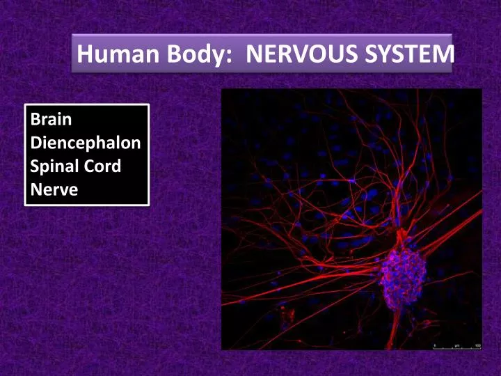
Human Body: NERVOUS SYSTEM. Brain Diencephalon Spinal Cord Nerve. Nervous system = master control and communication. Three functions:. 1. Gathering Sensory input (information about stimuli). Integration (interpretation and decision-making about the stimuli). !.

E N D
Human Body: NERVOUS SYSTEM BrainDiencephalonSpinal CordNerve
Nervous system = master control and communication Three functions: 1. Gathering Sensory input (information about stimuli). • Integration (interpretation and decision-making about the stimuli). ! 3. Effects a response – motor output. RUN AWAY!
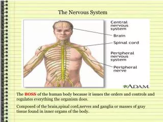
Organization of the Nervous System CNS:central nervous systemincludes brain and spinal cord PNS:peripheral nervous systemincludes nerves
Organization of the Nervous System PNS:peripheral nervous systemincludes nerves
Afferent (Sensory) Division:conveys impulses to the CNS Efferent (Motor) Division:conveys impulses from the CNS Subdivisions: Somatic/Voluntary NS Autonomic/Involuntary NS
Supporting Cells Supporting the CNS: Neuron = nerve cell Nervous Tissue
Nervous Tissue • Supporting the CNS: • Neuroglia (Glia) • nerve glue cells • Astrocytes: • type of glial cell • 50% of nerve tissue • protect neurons from • harmful substances in blood
Nervous Tissue Supporting the CNS: Ependymal cells on the surface of the choroid plexus secrete cerebrospinal fluid.
Supporting the CNS: Microglia found within the central nervous system are similar to macrophages. Nervous Tissue
Supporting the CNS: Extensions from the oligodendrocyte form the myelin sheaths of axons within the central nervous system. Nervous Tissue
Nervous Tissue Supporting the CNS: Extension from the Schwann cell forms a myelin sheath called the neurilemma around each axon within the peripheral nervous system.
Nervous Tissue Myelin sheath (blue) No Myelin sheath
Anatomy of a Nerve Cell Myelin Sheaths and Shingles shingleshelpline.com b4tea.com healthinplainenglish.com herpeszostervirus.com emed.com.au prlog.org
Nervous Tissue Supporting the CNS: Neuron cell bodies within ganglia are surrounded by satellite cells. Little is known of their function.
Anatomy of a Nerve Cell Myelin Sheath Node of Ranvier
Anatomy of a Nerve Cell Dendrites conduct impulses toward the cell body. Axons conduct impulses away from the cell body.
Anatomy of a Nerve Cell CNS clusters of nerve cells = nuclei PNS clusters of nerve cells = ganglia ganglion nuclei neuromedia.neurobio.ucla.edu eftlab.org
Anatomy of a Nerve Cell Tracts = bundles of nerves in CNS Nerves = bundles of nerves in the PNS Tract Nerves spiltmartini.com
Anatomy of a Nerve Cell Types of Sensory Receptors 1. Naked Nerve Endings = pain & temperature
Anatomy of a Nerve Cell Types of Sensory Receptors 2. Meissner’s corpuscles = touch kushtush.com ttouchnorth.co.uk
Anatomy of a Nerve Cell Types of Sensory Receptors 3. Pacinian corpuscle = deep pressure russiablog.org responsiblemarketing.com impactlab.net
Anatomy of a Nerve Cell • Types of Sensory Receptors • Proprioceptors = • muscular stretching or tension zimbio.com webmd.com
Anatomy of a Nerve Cell White matter = myelinated regions of the brain Gray matter = unmyelinated regions of the brain The Human Brain: How We Decide
Anatomy of a Nerve Cell White matter and gray matter in MRI scans: Comparing normal brain (left) with Huntington’s Diseased brain (right). radlink.com.sg wordadaywonder.com http://player.discoveryeducation.com/index.cfm?guidAssetId=B1721030-D95E-45BF-B764-D4AC4026D0C0&blnFromSearch=1&productcode=US
The Nerve Impulse • Major functional properties: • Irritability • …ability to respond to stimuli I am irritable! And…You are getting on my very last nerve!
The Nerve Impulse Conductivity…ability to conduct electrical currents • Resting membrane – • Na+ and K+ levels are equal Na+ Na+ Na+ Na+ Na+ Na+ K+ K+ K+ K+ K+ K+ Nerve impulses are “all-or-nothing” events.
Conductivity The Nerve Impulse • B. Depolarization starts – • Na+ moves into nerve cell. • Cell depolarizes (becomes too + inside)Action Potential is generated. Na+ Na+ Na+ Na+ Na+ Na+ Depolarization K+ K+ K+ K+ K+ K+ K+ ACTION POTENTIAL De = reversal
Conductivity The Nerve Impulse • C. Action potential is propagated. Na+ Na+ Na+ Na+ Na+ Na+ Depolarization K+ K+ K+ K+ K+ K+ K+ K+ K+ K+ K+ K+ K+ ACTION POTENTIAL
Conductivity The Nerve Impulse Re = again • C. REpolarization. REpolarization K+ Na+ K+ Na+ K+ Na+ K+ Na+ K+ Na+ K+ Na+ Back to equal! Too much + inside! Nerve Impulse http://highered.mcgraw-hill.com/sites/0072495855/student_view0/chapter14/animation__the_nerve_impulse.html
Nerve to Nerve Neurotransmitter_Synapse_3D_Animation
Stimulation of Muscles: • An action potential arrives at a presynaptic terminal. • The Calcium ion channel opens releasing calcium ions into the presynaptic terminal. • Calcium ions cause the synaptic vesicle to move to the synaptic cleft. • The synaptic vesicle releases ACH neurotransmitter into the cleft. • ACH diffuses across the cleft and and binds to ACH receptors on the muscle fiber membrane. • Sodium channels open and release sodium into the muscle. • The muscle membrane depolarizes and a postsynaptic action potential results. Ca+ Ca+ Ca+ Ca+ NA+ ACH NA+ NA+
Reflexes Auto = self Reflex = rapid, predictable, involuntary response ReflexArc = neural pathway of a reflex (goes only one way) Autonomic reflexes regulate smooth muscles
Reflexes Soma = body http://www.sumanasinc.com/webcontent/animations/content/reflexarcs2.html Somatic reflexes stimulates skeletal muscles
Brain Stem About the size of a thumb in diameter and about 3 inches long. rainbowrehab.com
Brain Stem Midbrain = small part that relays impulses and controls reflexes for vision and hearing.
Brain Stem Pons = bridge Pons = rounded structure of mostly fiber tracts involved in the control of breathing.
Brain Stem Medula Oblongata = merges into spinal cord; regulates vital internal activities including heart rate, blood pressure, breathing, swallowing, vomiting, etc.
Brain Stem Cerebellum = Outer gray matter; inner white matter. Provides timing for skeletal muscle activity, controls balance, and equilibrium. Compared to “autopilot” because it constantly checks and adjusts. Ataxia = Clumsy and disorganized movements as a result of damage to the cerebellum.
Sheep Brain Dissection Lab http://www.youtube.com/watch?v=08iZVEa5H9Y&feature=related
THE CNS The BRAIN http://www.youtube.com/watch?v=UFkSJemE4Pw Brain Function and Anatomy http://www.waiting.com/brainfunction.html
Brain Explorer An interactive resource for knowledge about the human brain. http://www.brainexplorer.org/brain_atlas/brainatlas_index.shtml Bon Voyage! Go Explore the Brain.
BRAIN FUN! RIGHT BRAIN VS LEFT BRAIN http://www.wherecreativitygoestoschool.com/vancouver/left_right/rb_test.htm http://homeworktips.about.com/library/brainquiz/bl_leftrightbrain_quiz.htm http://similarminds.com/brain.html http://frank.mtsu.edu/~studskl/hd/learn.html What are your results? Are you right or left brained? What does it mean?
Nervous System Protection of CNS Meninges = three membranes covering the CNS structures (p 211) Duramater = outermost layer Dura = hard Mater = mother netterimages.com schools-wikipedia.org
Arachnoid Mater = web-like middle layer commons.wikimedia.org Arachno = spider
Pia Mater = innermost layer following folds commons.wikimedia.org Sheep Brain Dissection http://www.youtube.com/watch?v=vE3Yf_xy_mE pia = gentle
Meningitis = inflammation of meninges http://video.about.com/infectiousdiseases/Meningitis.htm http://www.nmaus.org/programs/getting-it/ http://www.healthline.com/hgy-transcripts/meningitis-overview http://healthline.healthology.com/hybrid/hybrid-autodetect.aspx?content_id=2582&focus_handle=childrens-diseases&brand_name=healthline
Cerebrospinal Fluid (CSF) = fluid surrounding the brain and spinal cord Continually made from blood plasma in choroid plexuses which hang from the “roof” of the brains ventricles. Circulates continuously by being produced and then drained back into blood plasma to keep a constant volume of about 150 ml. Function: protection Spinal Tap = sampling technique to test CSF. Hydrocephalus = “water on the brain”
Keeps neurons separated from blood-borne substances. BLOOD-BRAIN BARRIER Made of LEAST PERMEABLE capillaries in the body. Can’t keep out fats, respiratory gases, alcohol, nicotine, or anesthetics. vandenberg.af.mil
Brain Injuries Traumatic Brain Injuries and Brain Dysfunctions Head injuries are the leading cause of accidental death in the USA. Concussion = slight injury, dizziness, brief loss of consciousness. http://www.pennmedicine.org/encyclopedia/em_DisplayAnimation.aspx?gcid=000034&ptid=17 Contusion = marked tissue destruction, coma Cerebral Edema = swelling of the brain, death