Download
1 / 26
270 likes | 455 Vues
Growth of EM Method for Determining Structures of Macromolecular Assemblies. What is cryo EM?. EM = (Transmission) Electron Microscopy Cryo EM = technique where biological samples are preserved in vitreous ice and imaged by EM at cryogenic temperatures.

E N D
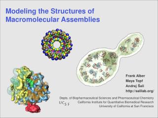
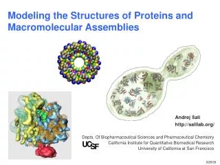
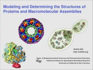
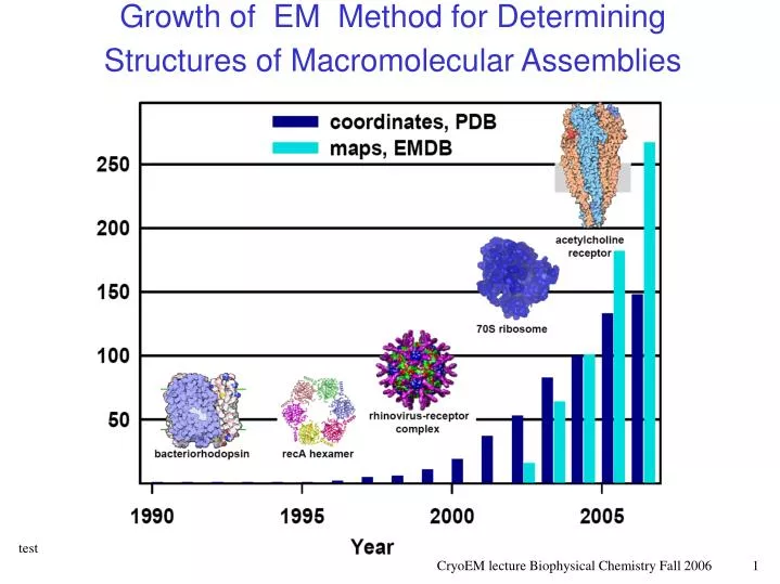
Growth of EM Method for Determining Structures of Macromolecular Assemblies
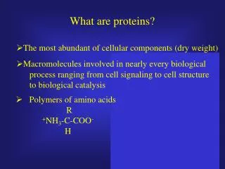
What is cryo EM? • EM = (Transmission) Electron Microscopy • Cryo EM = technique where biological samples are preserved in vitreous ice and imaged by EM at cryogenic temperatures. • EM reconstruction = 3D maps are generated by averaging over many EM images.
From Sample to Structure Slide from Wah Chiu, Baylor
Single Particles • MW lower limit: ~200 kDa Icosahedral virus Ribosome Image sources: EMDB, Joachim Frank, Sacha De Carlo
Filaments Acetylcholine receptor tubular crystal Tobacco Mosaic Virus gingi.uchicago.edu/achr.html Actin-myosin complex ami.scripps.edu
2D Crystals • Aquaporin 2D crystals, electron diffraction • Lipid-protein interactions Gonen et al. 2005. Nature 438:633-8.
Ensembles • Reconstruction of HIV capsids by cryoEM tomography Benjamin et al. 2005. J. Mol Biol. 346:577-588.
specimen preparation SUPPORT GRID 3.05 mm 400 divisons/inch 1 division/66 microns Copper or other metal Manufactured “holey carbon grid” Holes typically 2 microns diameter 2spi.com/catalog
High contrast image No special temperature control Essentially no radiation damage Particle distorted Image = stain “shell” around the particle Low resolution method: 20-15 Å Great choice for initial sample screening Low contrast image Sample maintained at cryogenic temperature (85 K) High radiation damage Particle undistorted Image is of the actual particle Higher resolution obtained: 15-4 Å Best choice for reconstruction Negative Stain vs. Vitreous Ice Specimen in Stain Cryogenic Specimen vitreous ice layer uranyl acetate
Imaging--TEM microscope Sample Goes here Swap out to view diffraction Swap out screen to record image Film/CCD camera cryoem.berkeley.edu/~nieder
Cryo EM Experiment Characteristics • Images must be taken with low electron doses to prevent radiation damage • Averaging over thousands of individual particles and/or “asymmetric units” Image: http://ami.scripps.edu
Data Collection and Initial Image Processing • Collect image set (20-100 images, vary focus) • Pick Particles (4000-100,000) • Perform contrast-transfer-function (CTF) correction for each image • Center, align, classify, make “class averages”
Particle Selection Raw image Auto-Select particles Particle Composite For 1 raw image
Recovering 3D from 2D figure from Joachim Frank
Reconstruction Cycle cryoem.berkeley.edu/~nieder Final map
Map Quality figure from Joachim Frank
Structure Analysis Methods to interpret cryo EM map volumes: • “segmentation” -- identifying different parts of the map • “fitting” --placing atomic coordinates into the map, e.g., from X-ray structures • very new: normal mode refinement to improve fit
Segmentation • herpes simplex capsid heterotrimer • Epsilon 15 bacteriophage Images from Wah Chiu
Fitting Rossmann, M. G. et al 2004. Curr Opin Struct Biol 14:171-80. Kostyuchenko, V. A. et al 2005 Nat Struct Mol Biol 12:810-3.
Archiving • wwpdb.org • Coordinates • www.ebi.ac.uk/msd/emdep • Maps • Soon…a single archive site for maps and models
References • The Definitive Textbook: • Frank, J. 2006. Three-dimensional electron microscopy of macromolecular assemblies : visualization of biological molecules in their native state. 2nd ed. New York : Oxford University Press. • Reviews: • Chiu, W., M. L. Baker, and S. C. Almo. 2006. Structural biology of cellular machines. Trends Cell Biol 16:144-50. • Nickell, S., C. Kofler, A. P. Leis, and W. Baumeister. 2006. A visual approach to proteomics. Nat Rev Mol Cell Biol 7:225-30.
Resources • Research-Resource Centers for Molecular Microscopy: www.ncrr.nih.gov/ncrrprog/btdir/Microsco.asp • EM software tools: en.wikipedia.org/wiki/Software_tools_for_molecular_microscopy • Visualization: www.cgl.ucsf.edu/chimera/ • EMDB map database: www.ebi.ac.uk/msd-srv/emsearch • Icosahedral Viruses: viperdb.scripps.edu
Cool Movies based on cryoEM data • Microtubules: http://cryoem.berkeley.edu/animations.shtml • T4 virus: www.nsf.gov/news/news_summ.jsp?cntn_id=100420&org=MCB&from=news • Ribosome: www.wadsworth.org/databank/electron • Actomyosin/kinesin: www.scripps.edu/cb/milligan/index.html T4 virus