Download
1 / 41
820 likes | 2.24k Vues
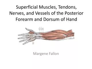
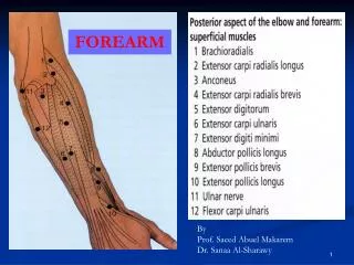
Muscles of the forearm. Posterior compartment. Post. Forearm. A: Extensor Digitorum B: Extensor Carpi Ulnaris C: Brachio Radialis D: Biceps Brachii E: Triceps F: Extensor Retinaculum. Muscles of the posterior compartment. Arranged In two layers: 1- A superficial layer

E N D
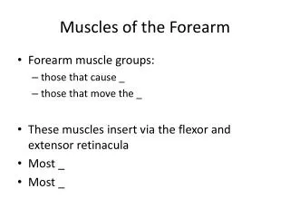
Muscles of the forearm Posterior compartment
Post. Forearm A: Extensor Digitorum B: Extensor Carpi Ulnaris C: BrachioRadialis D: Biceps Brachii E: Triceps F: Extensor Retinaculum
Muscles of the posterior compartment • Arranged In two layers: 1- Asuperficial layer 2- A deep layer.
Superficial Group • Brachioradialis • Extensor carpi radialis longus • Extensor carpi radialis brevis • Extensor digitorum • Extensor digiti minimi • Extensor carpi ulnaris • Anconeus • Common origin from the supracondylar ridge and lateral epicondyle of the humerus • Except for the brachioradialis and anconeus, the restextend as tendons into the hand.
Deep Group • Supinator • Abductor pollicis longus • Extensor pollicis brevis • Extensor pollicis Longus • Extensor indicis • Except for the supinator muscle, all these deep layer muscles originate from the posterior surfaces of the radius, ulna, and interosseous membrane and pass into the thumb and fingers.
Superficial Group Brachioradialis • Origin Lower 2/3 of the lateral supracondylar ridge of the humerus • Insertion Styloid process of the radius • Actions Elbow flexion in midprone position Pronation Supination • Innervation Radial nerve
Extensor Carpi RadialisLongus • Origin: • Lower 1/3 of lateral supracondylar ridge of humerus • Lateral epicondyle of humerus • Insertion: • Base of 2nd metacarpal (dorsal surface)
Extensor Carpi Radialis Longus • Action: • Extension of wrist • Abduction of wrist • Weak extension of elbow • Innervation: • Radial Nerve (C6,7)
Extensor Carpi RadialisBrevis • Origin: • Lateral epicondyle of humerus • Insertion: • Base of 3rd metacarpal (dorsal surface)
Extensor Carpi Radialis Brevis • Action: • Extension of wrist • Abduction of wrist • Weak extension of elbow • Innervation: • Radial nerve (C6,7)
Extensor Digitorum • Origin: • Lateral epicondyle of humerus • Insertion: • 4 tendons to bases of middle & distal phalanges of the 4 fingers
Extensor Digitorum • Action: • Extension of 2nd, 3rd, 4th, 5th phalanges • Extension of wrist • Weak extension of wrist • Innervation: • Radial nerve (C6,7,8)
Extensor DigitiMinimi • Origin: • Lateral epicondyle of humerus • Insertion: • Base of middle & distal phalanxes of 5th phalange (dorsal surface)
Extensor Digiti Minimi • Action: • Extension of little finger • Weak wrist extension • Innervation: • Radial nerve (C6,7,8)
Extensor Carpi Ulnaris • Origin: • Lateral epicondyle of humerus • Insertion: • Base of 5th metacarpal (dorsal side)
Extensor Carpi Ulnaris • Action: • Extension of wrist • Adduction of wrist • Weak extension of elbow • Innervation: • Radial nerve (C6,7,8)
Anconeus • Origin: origin from lateral epicondyle of humerus; • Insertion : At posterolateral surface of olecranon process of ulna • Action: Extension of forearm at elbow • Nerve supply: Radial nerve (C7, C8 and T1)
Deep Group Supinator • Origin: • from lateral surface of olecranon, • lateral epicondyle & • elbow ligaments • Insertion: • posterior and lateral surface of proximal 1/3 of radius • Actions: supination • Nerve Supply: Radial n.
Abductor PollicisLongus • Origin: • Posterior aspect of radius • Midshaft of ulna • Insertion: • Base of 1st metacarpal (dorsal surface)
Abductor Pollicis Longus • Action: • Abduction of thumb • Abduction of wrist • Innervation: • Radial nerve (C6, 7)
Extensor PollicisBrevis • Origin: • Posterior surface of lower middle radius • Insertion: • Base of proximal phalanx of thumb (dorsal surface)
Extensor Pollicis Brevis • Action: • Extension of thumb • Weak extension of wrist • Innervation: • Radial nerve (C6,7)
Extensor PollicisLongus • Origin: • Posterior lateral surface of lower middle ulna • Insertion: • Base of distal phalanx of thumb (dorsal surface)
Extensor Pollicis Longus • Action: • Extension of thumb • Extension of wrist • Innervation: • Radial nerve (C6,7,8)
Extensor Indicis • Origin: • Middle to distal 1/3 of posterior ulna • Insertion: • Base of middle & distal phalanxes (dorsal surface)
Extensor Indicis • Action: • Extension of index finger • Weak wrist extension • Innervation: • Radial nerve (C6,7,8)
Blood supply of the posterior compartment 1-Radial artery 2-Posterior interosseous artery origin: common interosseous branch of the ulnar artery recurrent interosseous artery End by joining to dorsal carpal arch of the wrist 3-Anterior interosseous artery origin: common interosseous branch of the ulnar artery
POSTERIOR INTEROSSEOUS ARTERY Posterior interosseous artery • Branch of Common interosseous artery • Crosses over interosseousmemb to reach post compartment. • Gives recurrent interosseous artery, which takes part in anastomosis around elbow joint . • Passes between supinator and abductor pollicislongus to supply superficial extensors. • Terminates by joining dorsal carpal arch of wrist after receiving terminal end of anterior interosseous artery.
ANTERIOR INTEROSSEOUS ARTERY • Branch of the common interosseous artery. • Situated in the anterior compartment of the forearm on the interosseous membrane. • Perforating branches, which pass through the interosseous membrane to supply deep muscles of the posterior compartment
VEINS • Deep veins of the posterior compartment generally accompany the arteries. They ultimately drain into brachial veins associated with the brachial artery in the cubital fossa.
POSTERIOR INTEROSSEOUS NERVE • Branch of radial nerve • Enters back between sup. &deep fibres of supinator • Runs on post surface of interrosseousmemb up to wrist • Branches: A: Muscular B: Articular C: Sensory
EXTENSOR COMPARTMENT :NERVE SUPPLY • Anconeus • Brachioradialis • Extensor carpiradialislongus • Extensor carpiradialisbrevis • Extensor digitorum • Extensor digitiminimi • Supinator • Abductor pollicislongus Extensor pollicislongus • Extensor pollicisbrevis • Extensor indicis • Extensor carpiulnaris Aconeus Brachioradialis ECRL Radial nerve ECRB ED EDM ECU S APL EPB EPL EI Posterior interosseous nerve
APPLIED ANATOMY • WRIST DROP: Injury to radial nerve above the origin of post interosseous nerve • Paralysis of extensor muscles of forearm • If deep branch of radial nerve is damaged • Ext carpi radialislongus and brevis are spared wrist drop is absent
Muscles of the Forearm Post View Origin :Lateral epicondyle of humerusInsertion Lateral surface of olecranon andsuperior part of posterior surface of ulna Action Assists triceps in extending forearm;stabilizes elbow joint; abducts ulna during pronationInnervation Radial nerve (C7, C8 and T1) Anconeus Radial nerve innervates the BEST. BrachioradialisExtensorsSupinatorTriceps
Deep posterior compartment. • - abductor pollicis longus • - extensor pollicis brevis • - extensor pollicis longus • - extensor indicis • - supinator • Functional organization: • extend hand at wrist • extend / abduct thumb • extend index finger • supinate abductor pollucis longus extensor pollicis brevis extensor pollicis longus extensor indicis
Deep Dissection Forearm Post Ant
Quiz 12. Pronator Teres 13. Flexor carpiradialis: 14. Flexor Digitorum: 15. Extensor carpiulnaris: 16. Extensor digitorum: 17. Extensor carpiradialis:
Common Extensor Origin • Common Flexor Origin