Download
1 / 33
330 likes | 444 Vues
Transforming Pathology Teaching with Digital Technology. Nicholas Hardin, MD, Jill Jemison, Greg Sharp, MD, and Ted Bovill, MD. UVM College of Medicine: Snapshot. Established in 1822; 7 th oldest medical school Who 434 medical students; 25 MD/PhD students

E N D
Transforming Pathology Teachingwith Digital Technology Nicholas Hardin, MD, Jill Jemison, Greg Sharp, MD, and Ted Bovill, MD
UVM College of Medicine: Snapshot • Established in 1822; 7th oldest medical school • Who • 434 medical students; 25 MD/PhD students • Faculty: 79 basic science, 526 clinical, 1440 volunteer • Clinical sites in VT, FL, ME, CT • Technology • Dell Latitude Tablets • Blackboard LMS • SecurExam Browser • Learning Objects Suite • Polycom PVX • Homegrown Patient tracking, virtual microscopy, podcasting
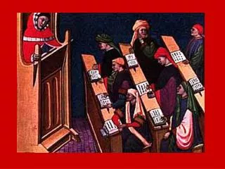
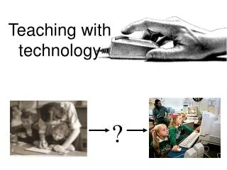
How Digital Technology has changed our teaching • Dark room to light • One person at a scope to entire group seeing same slide/discussing • Changing or inserting slide into lab much easier (one scanned vs. 120 copies) • Easier to share special stains, cytology, needle biopsies, unique specimens • Students asked for and got more videos
Feedback • “I thought that the virtual microscope was a great tool to study histology. I really liked being able to study and work with the slides where ever I was, and not having to cart around a microscope.”
Lessons learned • It takes money and time, so have high-level support • Plan and budget to scan more slides that you currently use • Faculty were initially against digital scope, but are now on board and enjoying it • We at UVM have only begun to tap the potential of these powerful tools
Acknowledgements • UVM COMET Lab: Andrew Verhelst: Digital Microscope, and Judith Kessler: Museum photo cleanup and programming • UVM Medical Photography: Gross Museum photography