Download
1 / 4
40 likes | 110 Vues
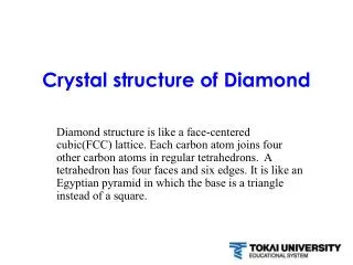
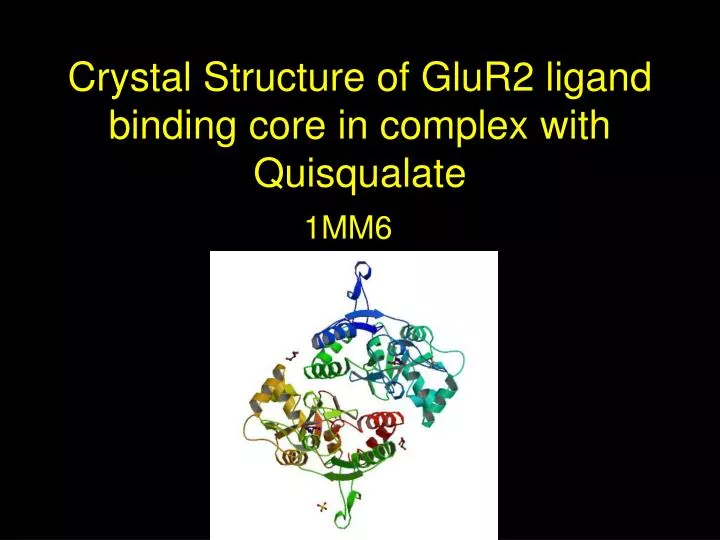
Crystal Structure of GluR2 ligand binding core in complex with Quisqualate. 1MM6.

E N D
Crystal Structure of GluR2 ligand binding core in complex with Quisqualate 1MM6
Glutamate receptor 2 is a protein encoded by the GRIA2 gene. It is the major excitatory neurotransmitter in the mammalian brain as it mediates most of the fast excitatory synaptic transmission in the mammalian central nervous system.
Quisqualate is a high-affinity receptor that elicits maximum peak current responses, and stabilizes the ligand binding core in a fully closed conformation In red are the QUS receptor sites.
The mechanism of quisqualate binding is different from that of AMPA but similar to that of glutamate • A comparison of the three complexes reveals distinct binding mechanisms, particularly in the region of a hydrophobic pocket • The hydrophobic pocket, plays an important role in determining receptor subtype specificity. The regions in blue are hydrophobic