Download
1 / 63
630 likes | 723 Vues
Heart Disease & Cancer. These diseases kill more people than all others combined (except for dying “naturally” of organ failure due to old age) ~ 247 heart disease deaths / 100,000 people / year (USA - 2001)* ~ 195 cancer deaths / 100,000 / year (USA - 2001)*

E N D
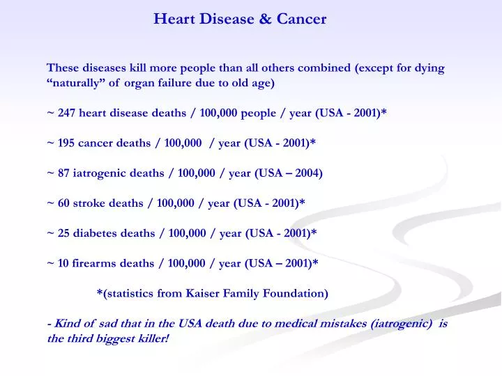
Heart Disease & Cancer These diseases kill more people than all others combined (except for dying “naturally” of organ failure due to old age) ~ 247 heart disease deaths / 100,000 people / year (USA - 2001)* ~ 195 cancer deaths / 100,000 / year (USA - 2001)* ~ 87 iatrogenic deaths / 100,000 / year (USA – 2004) ~ 60 stroke deaths / 100,000 / year (USA - 2001)* ~ 25 diabetes deaths / 100,000 / year (USA - 2001)* ~ 10 firearms deaths / 100,000 / year (USA – 2001)* *(statistics from Kaiser Family Foundation) - Kind of sad that in the USA death due to medical mistakes (iatrogenic) is the third biggest killer!
Coronary Artery Disease & Cancer Because there are many common lifestyle-associated risks for both CAD and Cancer, these two diseases are discussed together (more or less)
A very important question to ask is why would anyone suspect that these two diseases could be related to diet (and lifestyle) at all We all know that “cholesterol causes heart disease” and that “smoking causes heart disease and lung cancer” but these common associations may not be that simple…
If smoking was such an important component of risk for heart disease and lung cancer one would expect Japan to have far more heart disease and lung cancer than USA, Scotland or France, not so much less.
Atherosclerosis (CAD) is actually an inflammatory disease that is greatly affected by life-style issues. Research from different countries on the incidence of death due to heart disease in combination with various lifestyle factors illustrates this very nicely. For example, smoking cigarettes is strongly associated with risk for heart disease in the USA where nearly 40% of men smoke. In Japan, where about 70% of men smoke (1.75 x more smokers), there are almost 35% FEWER deaths (per 100,000) than in the USA. One might expect that with more smokers, there would be more heart disease but that is not the case. Somewhat similar results are observed when you compare France to the USA, although the incidence of smoking is almost the same in the two countries. Two major differences, however, are in fat consumption and cholesterol levels in serum. Paradoxically, cholesterol levels are much higher in France, as is saturated fat consumption. While USA (only) studies correlate cholesterol and fat levels with heart disease deaths due to heart disease are much lower in France (indicating no real correlation between disease and cholesterol or fat consumption. Some have called this a paradox. When the dietary and activity habits of people who live in these countries are taken into consideration however, then the apparent differences in heart disease (deaths) become understandable.
Consumption of fruits and vegetables as well as fish is much higher in France (and Japan) than in the USA. In addition, the amount of physical activity performed by the French and Japanese also is so much greater than that performed by Americans. These lifestyle differences can explain the large differences in heart disease among these countries; especially when you consider that heart disease (atherosclerosis) is predominantly a disease of chronic inflammation and not a disease of disordered lipids
Again, with so many more smokers, one would expect China to have so much more disease than Germany - although the lower-risk lipid profile may be part of why there is less heart disease deaths..
For a variety of cancers both diet & exercise contribute to a significant percentage of risk.
Even when looking at the major smoking-associated cancers, diet still is a major component of risk
Smokers appear to be very different people than non-smokers in terms of overall dietary habits: maybe that has something to do with the “smoking” risk...
For atherosclerosis, foam-cell production is the primary causal mechanism for initiating the deposition of plaque; note how 48 hours of no smoking or C + E supplements alter foam-cell production in cell culture experiments with smokers’ blood.
Some Summary Conclusions of Epidemiology Data The diet and lifestyle of smokers are very different from non-smokers High incidence of elevated serum cholesterol does not necessarily lead to a high incidence of heart disease Foam cell production in active smokers can be drastically reduced High incidence of smoking does not necessarily lead to a high incidence of lung cancer Various cancers have an attributable risk due to low vegetable consumption and low physical activity of anywhere from 30% to 50% (and quite possibly much more) To understand these complex relationships we really need to understand the mechanisms of the diseases We’ll start off with CAD
Atherosclerosis is actually an inflammatory disease of the coronary blood vessels
Smooth muscle cells, vascular endothelial cells, monocytes, and platelets are the major cells involved in the atherosclerotic process with oxidized lipoproteins and other oxidized lipids also being intimately involved.
The activation of proinflammatory and prohypertrophy signaling by mechanical disturbances of vascular cells due to low oscillating sheer forces and turbulent flow creates a sensitive proinflammatory environment within the cells located at bifurcates and inside curves of arteries. The initial infiltration of inflammatory cells contributes to the hypertrophy-adaptation response to ensure adequate downstream flow, as well as to the formation of Type I and II “fatty lesions”. Poor diet, overeating, and inadequate physical activity lead to poor redox control in cells, increased formation of AGEs, increased oxidation of HDL/LDL/CHOL, increased blood pressure, and greatly enhanced PRR activation and proinflammatory signaling; greatly increasing the risk for progression to the potentially fatal Type V and VI lesions.
As mentioned in the previous slide, proinflammatory signaling is initiated as a result of mechanical stimulation of the endothelial cells of the coronary blood vessels by oscillating sheer forces and turbulent flow. These forces are indicative of compromised flow and a normal response would be to initiate a hypertrophy/angiogenesis response in the blood vessel to increase the vessel wall thickness and enhance local pressure to increase flow.
Inflammatory signals result in the recruitment of monocytes into the blood vessel walls (between the endothelial cell layer and the smooth muscle cell layer). These monocytes mature into macrophages and they phagocytize any AGEs, oxidized lipids, and oxidized lipoproteins. These compounds bind to scavenger receptors on the macrophages cell membrane which then activate proinflammatory signaling. Another name for scavenger receptor is DAMP receptor. Thus scavenging oxidized and damaged molecules (AGEs) is a normal function of the macrophages; a function that activates the formation of phagolysosomes and ROS production. This of course, increases oxidative damage in the local area… When these macrophages scavenge large amounts of oxidized lipids and other damaged molecules they can actually get so large that they cannot get out of the vessel wall and they eventually die. When this happens, the lipids and fragments of cellular debris are left behind. If enough macrophages accumulate in one area of the blood vessel wall, consume sufficient AGEs and lipids and then die then lipids and cellular debris will build up in the blood vessel wall and cause it to bulge into the vessel lumen. Obviously this will impede blood flow. Unfortunately, impeded blood flow in the one spot will vastly increase both turbulence and the oscillations in sheer forces and therefor accelerate the processes of inflammation in this region and increase the infiltration and activation of macrophages. As a result of the chronic production of inflammatory signals there will be smooth muscle cell hyperplasia as well as fibrosis. Both of these processes are hallmarks of CAD and they can happen only as a result of inflammatory signals. The fatty plaque that builds up in the blood vessel wall contains both “extra” smooth muscle cells and fibrous tissue.
Monocytes adhere to the local area and bury beneath the endothelial layer where they differentiate into macrophages... and are then stimulated to ingest foreign particles as well as any oxidized lipids; as are any resident macrophages already in the vessel wall... platelets start to accumulate in the injured area as well...
Macrophages engulf advanced glycation end products, oxidized lipids, any oxidized lipoproteins, and transform into foam cells; releasing even more inflammatory cytokines
Foam cells accumulate and die in the extracellular space leaving behind their necrotic components and lipids while hyperplasia of smooth muscles (caused by the various growth factors produced as a result of the inflammatory signaling process) and accumulation of connective tissue form atherosclerotic lesions
With continued production of inflammatory signals there will be continual growth of the plaque and greater obstruction of the blood flow leading to greater deterioration of the endothelial cell layer. This creates an attractive rough area for platelets to adhere to in order to stimulate the local deposition of collagen and fibrin, leading to the development of a fibrous “cap”. Continual growth of the plaque can lead to complete occlusion of blood flow. As a result of the infiltrating platelets being activated they can release a variety of factors that destabilize the plaque increasing the likelihood that a piece of the plaque can break off and become a thrombus. A thrombus is a solid particle that can lodge in a smaller blood vessel to cause an infarct in some other tissue (brain or another area of the heart, for example) and death could possibly result - not necessarily a good thing.
As mentioned previously proinflammatory signaling is initiated as a result of mechanical stimulation of the endothelial cells of the coronary blood vessels by oscillating sheer forces and turbulent flow. These signaling events can be exaggerated by a variety of factors: • open-chain glucose / AGEs – elevated with insulin resistance/diabetes due to poor diet and inactivity • oxidized lipoproteins – elevated with poor antioxidant control/enhanced ROS due to poor diet and inactivity • oxidized lipids – elevated with poor antioxidant control/enhanced ROS due to poor diet and inactivity • and the exaggerated oscillations in sheer forces due to high blood pressure – elevated with obesity due to poor diet and inactivity • circulating proinflammatory signaling molecules that arise from other tissues – elevated with obesity due to poor diet and inactivity • Logically, the levels of any “toxic” molecules will be the same throughout the entire cardiovascular system while the blood pressure and sheer forces mill vary at different locations. Some of the highest blood pressures exist in the coronary vessels and the greatest oscillations in sheer forces exist at the bifurcations of the coronary vessels – the specific locations where atherosclerosis occurs.
SUMMARY CONCEPT OF THE CAUSE OF CAD CAD is caused by oscillating sheer forces and turbulent flow and the normal proinflammatory/hypertrophy signaling responses to these. These processes are exaggerated by poor antioxidant control that leads to enhanced ROS damage and to exaggerated signal transduction activities that leads to additional proinflammatory signaling arising from resident macrophages that are further activated via their normal scavenging functions. Poor antioxidant control and excess proinflammatory signaling are due primarily to poor diet and inactivity that results in insulin resistance, adipose tissue-gain, and hypertension. SUMMARY CONCEPT FOR PREVENTION By targeting the actual “causal” mechanisms of the disease, it is possible to attenuate the disease process and thereby greatly reduce risk for the disease.
A variety of phytochemicals are known to interfere with different components of the signaling pathways that lead to synthesis of the proinflammatory cytokines and eicosanoids.
By inhibiting production of both proinflammatory cytokines and eicosanoids, inflammatory signaling will be attenuated which will reduce the activation of monocytes and macrophages as well as reduce the recruitment of monocytes into the vessel wall.
Synthesis of the various antioxidant and redox control enzymes and compounds can be enhanced to optimize antioxidant and redox control. These enhanced protective effects are mediated by binding of the Nrf2 transcription factor to the antioxidant (electrophile) response element along with the small nuclear Maf proteins.
From all of this dietary and mechanistic information one can conclude that: consumption of a large variety of fresh fruits and vegetables, omega-3 fatty acids from cold water fish, alcohol, esp. red wine (in moderation) combined with regular stressful physical activity greatly reduces risk for heart disease such that “high risk” behaviors such as smoking may pose little or no risk.
Based on the mechanistic evidence atherosclerosis is NOT a disease of “abnormal” serum lipids, it is an INFLAMMATORY DISEASE (initiated by normal cellular responses to inadequate blood flow) & greatly exacerbated by DIETARY & EXERCISE INADEQUACIES
Based on the epidemiological evidence, cancer also has a very important dietary source of risk and (kinda like with CAD) only by understanding the mechanisms of cancer can we understand the complex relationship between diet and cancer
So, what exactly is cancer? In essence, cancer is simply uncontrolled cell division. The terms neoplasm and tumor are often used to indicate a cancerous growth; ie: neoplasm ~ tumor ~ cancer. Cancer comes in two types: Benign and Malignant. The process of developing cancer is generally called Carcinogenesis while the overall process is divided into three phases: Initiation: Oxygen radicals or chemical radicals form adducts with DNA bases of Stem Cells... unrepaired DNA damage leads to mutations in daughter cells following mitosis (well, there are other sources of mutations but these will suffice to start). Promotion: Accelerated cell division leads to the accumulation of more mutations (less time to repair DNA) and clonal expansion of the mutated cells. Progression: Accumulation of “appropriate” mutations leads to unrestrained cell division... acquisition of additional “appropriate mutations” leads to malignancy.
Thus, there appear to be two basic processes responsible for the carcinogenesis process: ROS- or Chemical radical-mediated DNA Damage (caused by mutagens: anything which causes DNA damage which results in mutations; of course repair infidelity or duplication errors also can do this ie. cause mutations) or, Enhanced rates of Cell Division to fix and accumulate DNA mutations; eventually leading to tumorigenesis (caused by mitogens; anything which results in abnormally high rates of cell division resulting in enhanced risk for cancer) Normal, controlled cell division is thus turned into mutated, uncontrolled cell division.
Cancer cells continuously divide in an uncontrolled fashion “Initiation” “Promotion” Normal cells enter the cell cycle only when stimulated by “well-controlled” growth signals. “Progression”
Obviously DNA damage is really important for causing mutations and cancer. So, the big question is: How is DNA damage caused? OXYGEN RADICALS CHEMICAL RADICALS (ROS) (carcinogens) Errors inherent in the process of Repair & DNA replication are also very important in producing mutations
Some Endogenous Sources of Reactive Oxygen Species - Cytochrome P450 (CYP) autooxidation - Xanthine oxidase - Ubiquinone and NADH oxidase in mitochondria - NADPH oxidase of phagocytic cells Some Sources of Chemical Radicals (aka carcinogens) - Cytochrome P450 (CYP) activation of xenobiotics We’ll deal with chemicalcarcinogens first
For the purposes of this discussion, xenobiotics refers to any compound not made by our tissues. Those which are not useful must be excreted. Fat soluble xenobiotics are usually metabolized to water-soluble products for excretion and it is this process which results in the formation of both ROS and carcinogens.
Different types of enzymes perform the chemical reactions necessary for these processes with CYP enzymes predominating in the activation of xenobiotics to carcinogens
Essentially, CYP enzymes take oxygen, reduce it, and the oxygen radical reacts with the xenobiotic to hydroxylate it concomitant with the release of water. If CYP enzymes do not work properly (uncoupled) they release ROS
Benzo(a)pyrene is a common xenobiotic found in smoke from burning plants & is a major carcinogen in tobacco smoke.
NNK is a major carcinogen found predominantly in tobacco smoke.
If all the adducts are added up one finds some interesting results: The majority of DNA damage to P53 – even in smokers – comes from ROS-mediated processes.
Events which alter the cell division cycle also are very important in carcinogenesis Stem cells are the predominant source of tumor cells.
Tissue Regeneration The Predominant Site of Tumorigenesis Under normal conditions Stem cells (SC) divide slowly to replenish the population of faster-dividing, short-term progenitor cells (PC). When progenitor cells divide, the displacement of their daughter cells toward the tissue environment forces cell:cell contact which, in concert with TGF and other factors, induces differentiation into mature tissue cells. These processes are under tight control by a variety of cell-signaling factors that modify activity of the various signal-transduction pathways
Regulation of Tissue Regeneration Elevated PGE2 and other growth factors activate various MAPK signal-transduction pathways in Stem Cells to initiate the activation and synthesis of a variety of proteins that are necessary for activating growth of the stem cell and the synthesis of DNA to duplicate the chromosomes in order to proceed to the mitosis phase. A decline in the levels of PGE2 and of growth factors induces G0 arrest via reducing MAPK activities. Progenitor cells divide more-or-less continually in response to normal levels of the same stress-response signaling with the displaced daughter cells differentiating into tissue cells.
ERK-MAPK - p38 MAPK - JNK-MAPK - PI3K/Akt The major signal transduction pathways (STPs) involved in regulating the growth and development of stem cells and progenitor cells are the ERK-MAPK, p38-MAPK, JNK-Mapk, PI3K/Akt, and the “Nfκβ” pathways. These STPs regulate synthesis of many different proteins; including those that are necessary for the different phases of cell division. If any of the genes that code for the various proteins of the STPs acquire an enabling mutation that renders them constitutively active then the constant activity of the associated STP might enhance risk for increasing rates of cell division and subsequently: tumorigenesis. Such a mutation would be called a DRIVER MUTATION.
Mutations in any of the genes that code for the various proteins that are involved in regulating cell division are called driver mutations: i.e.mutations that affect one or another function of dividing cells that increases the likelihood of them becoming tumor cells. Enabling mutations in any of the genes that code for proteins that stimulate different aspects of the cell division cycle and disabling mutations in any of the genes that code for proteins that block entry into cell division will enhance proliferative activity. Disabling mutations in the genes of any of the proteins involved in activating cell-cycle arrest, DNA repair, and apoptosis will not only enhance proliferative activity but greatly enhance the likelihood of mutations due to DNA damage.
DNA damage is central to the process of tumorigenesis. There is no shortage of oxidizing and alkylating species that are capable of reacting with DNA molecules t alter their structure and render them unrecognizable to the DNA polymerases; resulting in a variety of mutations produced during the S-phase. In addition to the types of mutations caused by oxidation events and alkylation events: predominantly transitions (A↔G, C↔T) and transversions (A↔C, T↔G); a variety of other consequences of DNA damage can occur: ∙OH can abstract a hydrogen from C5 of deoxyribose to leave a carbon-centered allyl radical which reacts with O2 to produce an oxyl-radical; leading to single strand breaks. When single-strand breaks are not repaired, they often lead to double-strand breaks during the S-phase; resulting in a variety of chromosomal mutations formed during mitosis. DNA:DNA and DNA:protein crosslinks also occur and these lead to chromosomal mutations as well.