Download
1 / 54
640 likes | 1.65k Vues
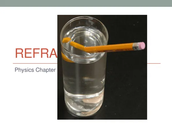
Refraction. Dr. Edia Asmara Soelendro, SpM Dr. Pandji A. Akbar, SpM. Refraction. Consists of : General Optics The optical system of the eye Clinical anomalies : refractive errors. Optic. Dioptri (D) : Lens power unit, is an inverse of focal distance in meters D = 1/f

E N D
Refraction Dr. Edia Asmara Soelendro, SpM Dr. Pandji A. Akbar, SpM
Refraction • Consists of : • General Optics • The optical system of the eye • Clinical anomalies : refractive errors
Optic • Dioptri (D) : Lens power unit, is an inverse of focal distance in meters D = 1/f • 1 D lens, parallel light will be directed into focal spot in 1 meter distance 2 D = 1/f ----> f = ? If f = 25 cm , ----> D = ?
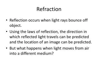
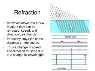
Parallel rays will be converged to the focus ---> Plus lens (+) • or will be diverged as if it comes from the focus ----> Minus Lens (-)
Principles • Rays coming from distance > 5 m parallel rays • Rays coming from distance < 5m divergent rays
Spherical lens • Is a lens with the same curvature diameter in all meridians Spherical Convex (+) Spherical Concave (-)
A convex lens may be regarded as a series of prisms bases toward the middle of the lens • A concave lens may be regarded as a series of prisms apex toward the middle of the lens
Prismatic Effect that occur on eye glasses explain : • Against motion with (+) Lens • With motion, with (-) Lens • Spherical Lens : • Plus sphere : Convex • characteristic : makes larger and nearer images 0 +4 +2 +2 +5 -1 Biconvex Plano K Concave K
Minus sphere : Concave • Characteristic : makes smaller and farther images Bi Concave Plano K Convex K • Parallel rays will be centered or diverged from the focus 0 -4 +1 -5 -2 -2
Cylindrical Lens • Is a kind of lens that have two meridians that are perpendicular to each other • The meridian that has no power is called the axis • The other meridian, has the power
Spherocylindrical Lens • Is a combination between spherical lens and cylindrical lens • Example : • S + 2.00 D C + 1.00 D X 90 0 0.00 + 2.00 + 2.00 0.00 + + 2.00 + 1.00 + 2.00 + 1.00 + 2.00 + 3.00
0 0 • Transposition • Methods : • Sphere : Sum with algebra ways SPH + CYL • Cylinder : replace power marks (Neg Pos), axis change 90 degrees • Example : S + 2.00 C + 1.00 X 90 S + 3.00 C - 1.00 X 180
Eye as an Optical Instrument • Refraction media : • Cornea n = 1.33 • Humour Aqueous n = 1.33 • Lens n = 1,41 • Vitreous body n = 1.33 • Haziness on refraction media --> disturbances of vision
Power of refraction of the eye ball • Totally : 60 dioptri • Cornea : 40 dioptri • Lens : 20 dioptri
Accommodation Process • Capability of adding the refraction power of the eye, by increasing the convexity of the lens • normal : rays that come from > 5 m - distance object regarded as parallel light; the eyes are in relax position, the images are focused right on the retina (fovea centralis)
For object at less than 5 meters distance, the rays do not come parallel but divergent. If the eyes are still in relax position, the images will be focused behind the retina. So the object will be seen blurred. These images must be moved forward so it will be focused on the retina by increasing the convexity of the lens. This process is called accommodation process.
This accommodation process happens as a result from the contraction of M. ciliaris in the ciliary body
These reflexes also happen during the accommodation process : • Accommodation • Miosis • Convergents Near Reflex
Refraction Anomalies • Normal : Emetropia • Anomalies : (ametropia) • Myopia • Hypermetropia • Astigmatism • Presbiopia
Emmetropia • Is the condition when the parallel rays focused exactly on the retina of the eye in relax condition ---> the visual acuity is maximum
Ametropia • Is the condition when the parallel rays are not focused exactly on the retina of the eye in relax condition. • The focal point may be behind or in front of the retina Hal 47, 4.2 Duke Elder
Myopia • Refractive condition in which, with accommodation completely relaxed, parallel rays are brought to a focus in front of the retina. • Myopic eye : refractive state over plus power
Factors that causing myopia : • Axial : The antero-posterior axis of the eye ball > normal • in this case, the refraction power of the cornea, lens and the lens position are normal. The eye usually looks like proptosis • Curvature : • The size of the eye ball ---> normal, but there is a increasing of the cornea/lens curvature • The change of the lens e.g. : intumescens cataract • Increasing of the refraction index • could occur on Diabetic patient • Changes of the lens location • changes of the lens position to the anterior after glaucoma surgery • lens subluxation
Clinical findings : • Farsightedness are blurred, nearsightedness are normal • Asthenopia • On high myopia : hemeralopia occurred caused by periphery retinal degeneration • Floating spots visualization caused by vitreous degeneration • screw up the eye lids together, in order to get a better vision • On high myopia ----> proptosis simulation, deep Anterior Chamber
Funduscopy : Tigroid fundus ---> thin retina and the choroid, myopic crescent arround the papilla area, sthaphyloma posterior
Complication : • Commonly occurred on high myopia 1. Degenarated and liquefied vitreous 2. Retinal detachment 3. Pigmentation changes + Macular bleeding 4. Strabismus • Myopia classification : • < 3.00 D = low myopia • 3.00 - 6.00 D = moderate myopia • > 6.00 D = high myopia/gravis
Treatment : • Low and moderate myopia : full correction with weakest spherical lens that give the best visual acuity • Example : VOD = 5/60 S -2.50 D = 6/7 S -2.75 D = 6/6 S -3.00 D = 6/6 S -3.25 D = 6/7 The glasses are S - 2.75 D • On high myopia, usually full correction are not given due to headache that may occurred. If necessary, reading glasses can be given ---> bifocal glasses
Prognosis : • Simplex/stationer, after puberty will be constant • Progressive myopia, the myopia will be continuously higher and complication may occurred
Hypermetropia • Is a refraction anomaly that without accommodation parallel rays will be focused behind the retina • Divergent rays from near object, will be focused farther behind the retina
Etiology : • Axial ---> eye ball diameter < N • Deminished convexity of cornea/lens curvature • Decreasing Refractive index • Changed lens position
Clinical manifestation : • H. Manifest ---> is detected without paralazing accommodation and is represented by the strongest convex glass needed , the patient sees most distinctly. It correspons to the amount of accommodation which he relaxes when a convex lens is placed before the eye. Devided into two types : • Facultative : Can be overcome by an effort of accommodation • Absolute : Can not be overcome
Total Hipermetrop : detected after the accommodation has been paralyzed with cylcopegic agents • Latent Hypermetrop : is the diference of the total hypermetrop with the manifest hypermetrop
Hypermetrop Latent Hypermetrop Hypermetrop manifest
Clinical finding : • Nearsightness are blurred • High hypermetropia at old age : farsightedness also blurred • Astenophia accommodative (eye strain) • Children : high hypermetropia usually occurring convergent strabismus (convergent squint)
Treatment : • If foria/tropia not present, apply strongest positive spherical lens that give the best visual acuity • If foria/tropia present, total hypermetrop correction. If necessary : bifocal eye glasses
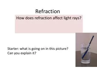
astigmatism • Refractive condition of the eye in which there is a difference in degree of refraction in diferent meridian, each will focused parallel rays at a different point. The shape of the images : • Line, oval, circle, never a point
Manifestation : • Regular astigmatism • Difference in the degree of refraction in every meredian. • Two principles meridian : • Maximmum refraction • Minimum refraction • Irregular astigmatism • Difference in refraction not only in different meridians, but also in different parts of the same meridian. Right angle to each other
Etiology of astigmatism : • Corneal curvature disturbances ---> 90% • Lens curvature disturbances ---> 10% • Type of Astigmatism : • Ast. M. Simplex C-2.00 X 90 • Ast. H. Simplex C+2.00 X 45 • Ast. M Compositium S-1.50 C-1.00 X 60 • Ast. H Compositium S+3.00 C+2.00 X 30 • Ast. Mixtus S+2.00 C-5.00 X 180 0 0 0 0 0
Ast. H. Simplex Ast. M. Simplex Ast. M Compositium Ast. H Compositium Ast. Mixtus
Presbiopia • Physiological changes because accommodation capability is lowering at old age Accommodation 16 10 6 2 Age 10 20 40 50 60
Presbiopia correction : • 40 years old S + 1.00 D • 45 years old S + 1.50 D • 50 years old S + 2.00 D • 55 years old S + 2.50 D • 60 years old S + 3.00 D • Consider the type of previous/history work • Tailor • Architect • Weld engineer
Refraction Examination Technique • Subjective : • Snellen chart/projector, alphabet , inverse E, picture, Landolt ring • Trial lens • Trial frame • Objective : • Children, incooperative, difficult correction, strabismus : • Ophthlamoscopy • Retinoscopy • Refractometer
Subjective • Check firstly just one eye : OD • Distance : 5 or 6 meters • VOD : …...(basic right eye visus) a. Trial and error • apply S + 0.50, better visus , add S+ until visus = 6/6 • S +0.50, lower visus, change to S -, increase S - until visus = 6/6 • S +/- not working ----> cylindrical • With astigmatism dial, stenoplic slit, cross cylinder • astigmatism dial : • Blurred line ----> C negative lens axis
One by one fogging • S + sp. Lens --> blurred vision, step by step distracting ---> best sp. • Nearsightedness/read • Both eyes at one time at required distance : use jaeger chart
ODS 6/6 headache, eye strain • Example : I. AVOD 2/60 S - 3.50 = 6/6 AVOS 3/60 S - 3.00 = 6/6 II.AVOD 2/60 S - 3.00 = 6/7 AVOS 3/60 S - 2.75 = 6/7 read ADD S + 1.50 Give Eye Glasses according to II ODS 6/6 w/o headache, eye strain
Objective • Use cyclopegic 1. Ophthlamoscopy : papilla clearly seen with which lens 2. Retinoscopy : • Ordinary ---> light source outside • streak -----> light source inside 3. Refactometer • Computerized • Lensmeter principal
Ideally : • Subjective • Objective with cyclopegic • Subjective once more without cyclopegic • Lens meter • Measuring lens power • Measuring focus distance
Measuring Pupillary Distance • drop the flashlight light onto both eyes, the light is coming from in front of the patient, The patient look at the observer forehead or the light ----> measure the distance of light spot between OD and OS ----> as near pupil distance • Far distance : • add 2 mm ---> for pupil distance less than 60 mm. • Add 3 mm ---> for pupil distance more than 60 mm
Eye Glasses • Monofocal • Bifocal • Progressive • Eye Glasses Prescription, the components are : • Which eye (OD or OS) • Power of the lens ( + or - , Power, axis) • ADDE for reading • Pupil distance far/near • Name of the patient