Download
1 / 56
640 likes | 1.91k Vues
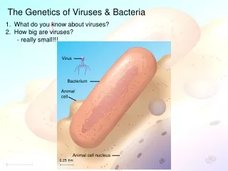
Genetics of Viruses and Bacteria. 0.5 m. Figure 18.1. Microbial Model Systems. Viruses called bacteriophages can infect and set in motion a genetic takeover of bacteria, such as Escherichia coli

E N D
0.5 m Figure 18.1 Microbial Model Systems • Viruses called bacteriophages can infect and set in motion a genetic takeover of bacteria, such as Escherichia coli • E. coli and phage model systems frequently use by researchers in studies that reveal broad biological principles • Viruses and bacteria have unique genetic mechanisms
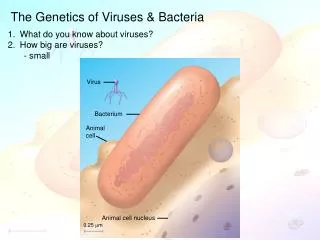
Virus Bacterium Animalcell 0.25 m Animal cell nucleus Characteristics of Viruses • Recall that bacteria are prokaryotes with cells much smaller and more simply organized than those of eukaryotes • Viruses are smaller and simpler still • Smallest viruses are only 20 nm in diameter • The virus particle, or virion, is just nucleic acid enclosed by a protein coat
Characteristics of Viruses • A virus has a genome but can reproduce only within a host cell • Scientists detected viruses indirectly long before they could see them • The story of how viruses were discovered begins in the late 1800s • Tobacco mosaic disease stunts growth of tobacco plants and gives their leaves a mosaic coloration • In the late 1800s, researchers hypothesized that a particle smaller than bacteria caused the disease • In 1935, Wendell Stanley confirmed this hypothesis by crystallizing the infectious particle, now known as tobacco mosaic virus (TMV)
Characteristics of Viruses • Viruses are very small infectious particles consisting of • Nucleic acid - genome • Protein coat which encloses the genome • And in some cases, a membranous envelope • Viral genomes may consist of • Double- or single-stranded DNA • Double- or single-stranded RNA
Capsomereof capsid RNA DNA Capsomere Glycoprotein 70–90 nm (diameter) 18 250 mm 20 nm 50 nm (b) Adenoviruses (a) Tobacco mosaic virus Capsids • A capsid is the protein shell that encloses the viral genome, it can have various structures • May be rod-shaped, polyhedral or complex • Composed of capsomeres – protein subunits; from one or a few types of proteins • Spikes or glycoproteins like the herpes shown
Membranous Envelope • Some viruses have envelopes which are membranous coverings derived from the membrane of the host cell • Maybe a single layer or double layer envelope • Bilipid bilayer with glycoproteins spikes protruding from the outer layer
Membranousenvelope Capsid RNA Glycoprotein 80–200 nm (diameter) 50 nm (c) Influenza viruses Membranous Envelope • Many animal viruses have a membranous envelope • The membrane cloaks the viral capsid, helps viruses infect their host • Derived from host cell membrane which is usually virus-modified • Viral glycoproteins on the envelope bind to specific receptor molecules on the surface of a host cell
Head DNA Tail sheath Tail fiber 80 225 nm 50 nm (d) Bacteriophage T4 Bacteriophages • Also called phages (T2, T4, T6) have the most complex capsids found among viruses • Icosohedral head encloses the genetic material; the protein tailpiece w/tail fibers attaches the phage to its bacterial host and injects its DNA into the bacterium
Viral Reproductive Cycles • Although a virus has a genome it can only reproduce within a host cell • Viruses are obligate intracellular parasites • Each virus has a host range - a limited number of host cells that it can infect • Recognize host cells by a complementary fit between external viral proteins and specific cell surface receptor sites • Viruses use enzymes, ribosomes, and small molecules of host cells to synthesize progeny viruses
VIRUS DNA Entry into cell and uncoating of DNA Capsid Transcription Replication HOST CELL Viral DNA mRNA Viral DNA Capsid proteins Self-assembly of new virus particles and their exit from cell Viral Reproduction
Reproductive Cycles of Phages • Phages are the best understood of all viruses • They through two alternative reproductive mechanisms: the lytic cycle and the lysogenic cycle • Lytic cycle - culminates in the death of the host • Lysogenic cycle - replicates the phage genome without destroying the host
1 Attachment. The T4 phage usesits tail fibers to bind to specificreceptor sites on the outer surface of an E. coli cell. 2 Entry of phage DNA and degradation of host DNA.The sheath of the tail contracts,injecting the phage DNA intothe cell and leaving an emptycapsid outside. The cell’sDNA is hydrolyzed. 5 Release. The phage directs productionof an enzyme that damages the bacterialcell wall, allowing fluid to enter. The cellswells and finally bursts, releasing 100 to 200 phage particles. Phage assembly 3 Synthesis of viral genomes and proteins. The phage DNAdirects production of phageproteins and copies of the phagegenome by host enzymes, usingcomponents within the cell. 4 Assembly. Three separate sets of proteinsself-assemble to form phage heads, tails,and tail fibers. The phage genome ispackaged inside the capsid as the head forms. Head Tail fibers Tails The Lytic Cycle • A phage reproductive cycle that culminates in the death of the host cell • Produces new phages and digests the host’s cell wall, releasing the progeny viruses • A phage that reproduces only by the lytic cycle is called a virulent phage • Bacteria have defenses against phages, including restriction enzymes that recognize and cut up certain phage DNA
Phage DNA The phage attaches to a host cell and injects its DNA. Many cell divisions produce a large population of bacteria infected with the prophage. Phage DNA circularizes Phage Occasionally, a prophage exits the bacterial chromosome, initiating a lytic cycle. Bacterial chromosome Lysogenic cycle Lytic cycle Certain factors determine whether The bacterium reproduces normally, copying the prophage and transmitting it to daughter cells. The cell lyses, releasing phages. Prophage Lytic cycle is induced Lysogenic cycle is entered or New phage DNA and proteins are synthesized and assembled into phages. Phage DNA integrates into the bacterial chromosome,becoming a prophage. The Lysogenic Cycle • The lysogenic cycle replicates the phage genome without destroying the host • The viral DNA molecule is incorporated by genetic recombination into the host cell’s chromosome • This integrated viral DNA is known as a prophage • Every time the host divides, it copies the phage DNA and passes it to the daughter cells • Phages that use both the lytic and lysogenic cycles are called temperate phages
Viral Classification • The nature of the genome is the basis for the common classification of animal viruses
3 patterns of viral replication • DNA DNA: If viral DNA is double-stranded, DNA replication resembles that of cellular DNA, and the virus uses DNA polymerase produced by the host. • RNA RNA: Since host cells lack the enzyme to copy RNA, most RNA viruses contain a gene that codes for RNA replicase, an enzyme that uses viral RNA as a template to produce complementary RNA. • RNA DNA RNA: Some RNA viruses encode reverse transcriptase, an enzyme that transcribes DNA from a RNA template.
Glycoprotein Viral envelope Capsid RNA(two identicalstrands) Reversetranscriptase RNA As Genetic Material - Retroviruses / Proviruses • The broadest variety of RNA genomes is found among the viruses that infect animals • Retroviruses, such as HIV, use the enzyme reverse transcriptase to copy their RNA genome into DNA • The viral DNA that is integrated into the host genome is called a provirus • Unlike a prophage, a provirus remains a permanent resident of the host cell
Glycoproteins on the viral envelope bind to specific receptor molecules(not shown) on the host cell, promoting viral entry into the cell. Capsid RNA 1 2 Envelope (with glycoproteins) Capsid and viral genome enter cell 3 HOST CELL The viral genome (red) functions as a template forsynthesis of complementary RNA strands (pink) by a viral enzyme. Viral genome (RNA) Template 5 Complementary RNA strands also function as mRNA, which is translated into both capsid proteins (in the cytosol)and glycoproteins for the viral envelope (in the ER). mRNA New copies of viral genome RNA are made using complementary RNA strands as templates. 4 Capsid proteins ER Copy of genome (RNA) Glyco- proteins 6 Vesicles transport envelope glycoproteins to the plasma membrane. 7 New virus 8 A capsid assembles around each viral genome molecule. The Reproductive Cycle Of An Enveloped RNA Virus • The host’s RNA polymerase transcribes the proviral DNA into RNA molecules • The RNA molecules function both as mRNA for synthesis of viral proteins and as genomes for new virus particles released from the cell
1 Reverse transcriptase catalyzes the synthesis of a DNA strand complementary to the viral RNA. 2 The virus fuses with the cell’s plasma membrane. The capsid proteins are removed, releasing the viral proteins and RNA. Membrane of white blood cell HIV Reverse transcriptase catalyzes the synthesis ofa second DNA strand complementary to the first. 3 Reverse transcriptase HOST CELL Viral RNA 4 The double-stranded DNA is incorporated as a provirus into the cell’s DNA. RNA-DNAhybrid 0.25 µm HIV entering a cell DNA ChromosomalDNA NUCLEUS Provirus 5 Proviral genes are transcribed into RNA molecules, which serve as genomes for the next viral generation and as mRNAs for translation into viral proteins. RNA genomefor the nextviral generation mRNA 6 The viral proteins include capsid proteins and reverse transcriptase (made in the cytosol) and envelope glycoproteins (made in the ER). 7 Capsids are assembled around viral genomes and reverse transcriptase molecules. Vesicles transport the glycoproteins from the ER to the cell’s plasma membrane. 8 9 New viruses bud off from the host cell. New HIV leaving a cell The Reproductive Cycle Of HIV, A Retrovirus
(b) The SARS-causing agent is a coronavirus like this one (colorized TEM), so named for the “corona” of glycoprotein spikes protruding from the envelope. (a) Young ballet students in Hong Kong wear face masks to protect themselves from the virus causing SARS. Viral Diseases in Animals • Viruses, viroids, and prions are formidable pathogens in animals and plants • Viruses may damage or kill cells by causing the release of hydrolytic enzymes from lysosomes • Some viruses cause infected cells to produce toxins that lead to disease symptoms • Emerging viruses are those that appear suddenly or suddenly come to the attention of medical scientists • Outbreaks of “new” viral diseases in humans are usually caused by existing viruses that expand their host territory • Severe acute respiratory syndrome (SARS) recently appeared in China
Bacterial Genetics • Rapid reproduction, mutation, and genetic recombination contribute to the genetic diversity of bacteria • Bacteria allow researchers to investigate molecular genetics in the simplest true organisms • The bacterial chromosome is usually a circular DNA molecule with few associated proteins • In addition to the chromosome, many bacteria have plasmids, smaller circular DNA molecules that can replicate independently of the bacterial chromosome
Mixture Mutantstrainarg+trp– Mutantstrainarg–trp+ RESULTS EXPERIMENT No colonies(control) No colonies(control) Researchers had two mutant strains, one that could make arginine but not tryptophan (arg+ trp–) and one that could make tryptophan but not arginine (arg trp+). Each mutant strain and a mixture of both strains were grown in a liquid medium containing all the required amino acids. Samples from each liquid culture were spread on plates containing a solution of glucose and inorganic salts (minimal medium), solidified with agar. CONCLUSION Mixture Only the samples from the mixed culture, contained cells that gave rise to colonies on minimal medium, which lacks amino acids. Mutantstrainarg+trp– Mutantstrainargtrp+ Coloniesgrew Because only cells that can make both arginine and tryptophan (arg+ trp+ cells) can grow into colonies on minimal medium, the lack of colonies on the two control plates showed that no further mutations had occurred restoring this ability to cells of the mutant strains. Thus, each cell from the mixture that formed a colony on the minimal medium must have acquired one or more genes from a cell of the other strain by genetic recombination. Mutation and Genetic Recombination • Since bacteria can reproduce rapidly new mutations can quickly increase a population’s genetic diversity • Further genetic diversity can arise by recombination of the DNA from two different bacterial cells
Mechanisms of Gene Transfer and Genetic Recombination in Bacteria • Three processes bring bacterial DNA from different individuals together • Transformation - Is the alteration of a bacterial cell’s genotype and phenotype by the uptake of naked, foreign DNA from the surrounding environment • Transduction - Phages carry bacterial genes from one host cell to another • Conjugation - Is the direct transfer of genetic material between bacterial cells that are temporarily joined
Mixture of heat-killed S cells and living R cells Heat-killed S cells (control) Living R cells (control) Living S cells (control) RESULTS Mouse dies Mouse healthy Mouse healthy Mouse dies Living S cells are found in blood sample Transformation • Transformation is the alteration of a bacterial cell’s genotype and phenotype by the uptake of naked, foreign DNA from the surrounding environment • For example, harmless Streptococcus pneumoniae bacteria can be transformed to pneumonia-causing cells
A+ A+ B+ B+ 2 Transduction Phage DNA 1 Phage infects bacterial cell that has alleles A+ and B+ Host DNA (brown) is fragmented, and phage DNA and proteins are made. This is the donor cell. Donorcell A bacterial DNA fragment (in this case a fragment withthe A+ allele) may be packaged in a phage capsid. 3 A+ Crossingover Phage with the A+ allele from the donor cell infects a recipient A–B– cell, and crossing over (recombination) between donor DNA (brown) and recipient DNA (green) occurs at two places (dotted lines). 4 A+ A– B– Recipientcell The genotype of the resulting recombinant cell (A+B–) differs from the genotypes of both the donor (A+B+) and the recipient (A–B–). 5 A+ B– Recombinant cell
Conjugation and Plasmids • Conjugation is the direct transfer of genetic material between bacterial cells that are temporarily joined • The transfer is one-way: One cell (“male”) donates DNA, and its “mate” (“female”) receives the genes • “Maleness,” the ability to form a sex pilus and donate DNA, results from an F (for fertility) factor as part of the chromosome or as a plasmid • Plasmids, including the F plasmid, are small, circular, self-replicating DNA molecules
A single strand of the F plasmid breaks at a specific point (tip of blue arrowhead) and begins tomove into the recipient cell. As transfer continues, the donor plasmid rotates(red arrow). DNA replication occurs inboth donor and recipientcells, using the single parental strands of the F plasmid as templates to synthesize complementary strands. The plasmid in the recipient cell circularizes. Transfer and replication result in a compete F plasmid in each cell. Thus, both cells are now F+. A cell carrying an F plasmid(an F+ cell) can form amating bridge with an F– celland transfer its F plasmid. 4 2 3 1 F plasmid Bacterial chromosome F+ cell F+ cell Mating bridge F– cell F+ cell Bacterial chromosome Conjunction and transfer of an F plasmid from and F+ donor to an F– recipient The F Plasmid and Conjugation • Cells containing the F plasmid, designated F+ cells, function as DNA donors during conjugation • F+ cells transfer DNA to an F recipient cell • Chromosomal genes can be transferred during conjugation when the donor cell’s F factor is integrated into the chromosome
Hfr cell F+ cell F factor 2 1 The circular F plasmid in an F+ cellcan be integrated into the circularchromosome by a single crossoverevent (dotted line). The resulting cell is called an Hfr cell (for High frequency of recombination). B+ D+ C+ C+ Hfr cell A+ D+ A+ B+ D+ A+ C+ B+ A+ B+ C– C– F– cell C– A+ B– D– A+ B– D– D+ B– D– C+ B+ A– A– A– C– The location and orientation of the F factor in the donor chromosome determine the sequence of gene transfer during conjugation. In this example, the transfer sequence for four genes is A-B-C-D. The mating bridgeusually breaks well before the entire chromosome andthe rest of the F factor are transferred. A single strand of the F factorbreaks and begins to move through the bridge. DNA replication occurs in both donor and recipient cells, resulting in double-stranded DNA B+ D– Since an Hfr cell has all the F-factor genes, it can form a mating bridge with an F– cell and transfer DNA. B– 3 4 6 5 A– A+ Temporary partial diploid Recombinant F– bacterium C– C– B+ B– D– D– B+ B– A– A– A+ A+ Two crossovers can result in the exchange of similar (homologous) genes between the transferred chromosome fragment (brown) and the recipient cell’s chromosome (green). The piece of DNA ending up outside thebacterial chromosome will eventually be degraded by the cell’s enzymes. The recipient cell now contains a new combination of genes but no F factor; it is a recombinant F– cell. 7 8 The F Plasmid and Conjugation • A cell with a built-in F factor is called an Hfr cell • The F factor of an Hfr cell brings some chromosomal DNA along when transferred to an F– cell • Thr transfer of part of the bacterial chromosome from an Hfr donor to an F– recipient results in recombination
R plasmids and Antibiotic Resistance • R plasmids confer resistance to various antibiotics • When a bacterial population is exposed to an antibiotic, individuals with the R plasmid will survive and increase in the overall population
Transposition of Genetic Elements • The DNA of a cell can also undergo recombination due to movement of transposable elements within the cell’s genome • Transposable elements: • Can move around within a cell’s genome • Are often called “jumping genes” • Contribute to genetic shuffling in bacteria
Insertion sequence 3 5 3 5 A T C C G G T… A C C G G A T… T A G G C C A … T G G C C T A … Transposase gene Inverted repeat Inverted repeat (a) Insertion sequences, the simplest transposable elements in bacteria, contain a single gene that encodes transposase, which catalyzes movement within the genome. The inverted repeats are backward, upside-down versions of each other; only a portion is shown. The inverted repeat sequence varies from one type of insertion sequence to another. Figure 18.19a Insertion Sequences • The simplest transposable elements, called insertion sequences, exist only in bacteria • An insertion sequence contains a single gene for transposase, an enzyme that catalyzes movement of the insertion sequence from one site to another within the genome
Transposon Antibioticresistance gene Insertion sequence Insertion sequence 5 3 5 3 Inverted repeats Transposase gene (b) Transposons contain one or more genes in addition to the transposase gene. In the transposon shown here, a gene for resistance to an antibiotic is located between twin insertion sequences. The gene for antibiotic resistance is carried along as part of the transposon when the transposon is inserted at a new site in the genome. Figure 18.19b Transposons • Transposable elements called transposons are longer and more complex than insertion sequences • In addition to DNA required for transposition, transposons have extra genes that “go along for the ride,” such as genes for antibiotic resistance
Control of Gene Expression • Every cell contains thousands of genes which code for proteins. • However, every gene is not actively producing proteins at all times. • To be expressed, a gene must be transcribed into m-RNA, the m-RNA must be translated into a protein, and the protein must become active. • Gene regulation can theoretically occur at any step in this process
Control of Gene Expression • Two categories of gene regulation: • Transcriptional controls - factors that regulate transcription • Post-transcriptional controls – factors that regulate any step in gene expression after transcription is complete • It is most efficient to regulate genes during transcription. • Both prokaryotes and eukaryotes rely primarily on transcriptional controls.
(a) Regulation of enzyme activity (b) Regulation of enzyme production Precursor Feedback inhibition Enzyme 1 Gene 1 Regulation of gene expression Enzyme 2 Gene 2 Gene 3 Enzyme 3 – Gene 4 Enzyme 4 – Gene 5 Enzyme 5 Tryptophan Regulating Prokaryotic Gene Expression • Prokaryotes can quickly turn genes on and off in response to environmental conditions. • This metabolic control occurs on two levels • Adjusting the activity of metabolic enzymes already present • Regulating the genes encoding the metabolic enzymes
Prokaryotic Gene Regulation • Response is facilitated by: • Simultaneous transcription and translation • Short-lived m-RNAs • Operons • Functionally related genes are often located next to each other and are transcribed as a unit. • For example E. coli, • 5 different enzymes are needed to synthesize the amino acid tryptophan • The genes that code for these enzymes are located together
Operons: The Basic Concept • In bacteria, genes are often clustered into operons, composed of • An operator, an “on-off” switch • A promoter • Genes for metabolic enzymes • An operon • Is usually turned “on” • Can be switched off by a protein called a repressor
trp operon Promoter Promoter Genes of operon RNA polymerase Start codon Stop codon trpR trpD trpC trpB trpE trpA DNA Operator Regulatory gene 3 mRNA 5 mRNA 5 Polypeptides that make up enzymes for tryptophan synthesis C E D B A Inactiverepressor Protein (a) Tryptophan absent, repressor inactive, operon on. RNA polymerase attaches to the DNA at the promoter and transcribes the operon’s genes. Figure 18.21a Prokaryotic Gene Regulation • A single promoter serves all 5 genes. (region where RNA polymerase binds to DNA and begins transcription) • The genes are transcribed as a unit, - one long mRNA molecule which contains the code to make all 5 enzymes
trp operon Promoter Promoter Genes of operon Start codon Stop codon RNA polymerase trpR trpD trpC trpB trpE trpA DNA Operator Regulatory gene 3 mRNA 5 mRNA 5 Polypeptides that make up enzymes for tryptophan synthesis C E D B A Inactiverepressor Protein (a) Tryptophan absent, repressor inactive, operon on. RNA polymerase attaches to the DNA at the promoter and transcribes the operon’s genes. Prokaryotic Gene Regulation • There is also a single regulatory switch, called the operator. • The operator is positioned within the promoter, or between the promoter and the protein coding genes. It controls access of RNA polymerase to the genes.
DNA No RNA made mRNA Active repressor Protein Tryptophan (corepressor) Tryptophan present, repressor active, operon off. As tryptophan accumulates, it inhibits its own production by activating the repressor protein. (b) Prokaryotic Gene Regulation • Transcription of the 5 coding genes in the tryptophan operon is blocked when a transcriptional repressorbinds to the operator. • The repressor binds to the operator only when there is a high level of tryptophan present:
Two Types of Negative Gene Regulation • In a repressible operon, binding of a specific repressor protein to the operator shuts off transcription • Repressible enzymes usually function in anabolic pathways • In an inducible operon, binding of an inducer to a repressor inactivates the repressor and turns on transcription • Inducible enzymes usually function in catabolic pathways
Promoter Regulatorygene Operator DNA lacl lacZ NoRNAmade RNApolymerase 3 mRNA 5 Activerepressor Protein (a) Prokaryotic Gene Regulation: Inducible Operon • The lactose operon in E. coli is an inducible operon • It controls the production of 3 enzymes needed to digest lactose (catabolism of a disaccharide made of glucose and galactose) • When lactose is absent, the repressor is active and the operon is off. • The lac repressor is innately active, and in the absence of lactose it switches off the operon by binding to the operator.
DNA lacl lacz lacY lacA lac operon RNApolymerase 3 mRNA 5 mRNA 5 mRNA 5' -Galactosidase Permease Transacetylase Protein Inactiverepressor Allolactose(inducer) lac Operon • If lactose is present, the repressor is inactivated and the operon is on • Allolactose, an isomer of lactose, turns on the operon by inactivating the repressor. In this way, the enzymes for lactose utilization are induced.
Positive Gene Regulation • Regulation of both the trp and lac operons involves the negative control of genes, because the operons are switched off by the active form of the repressor protein • Some operons are also subject to positive control via a stimulatory activator protein, such as catabolite activator protein (CAP) • CAP (catabolite activator protein) stimulates transcription of genes that allow E. coli to use other food sources when glucose is not present such as lactose
Operator RNA polymerase can bindand transcribe Promoter DNA lacl lacZ CAP-binding site ActiveCAP cAMP Inactive lac repressor InactiveCAP (a) Lactose present, glucose scarce (cAMP level high): abundant lac mRNA synthesized.If glucose is scarce, the high level of cAMP activates CAP, and the lac operon produces large amounts of mRNA for the lactose pathway. Positive Transcriptional Control • In E. coli, when glucose is scarce, the lac operon is activated by the binding of a regulatory protein, catabolite activator protein (CAP) • Low levels of glucose lead to high levels of cAMP • cAMP binds to CAP, CAP binds to CAP binding site, and transcription of lac mRNA is stimulated for catabolism of lactose
Promoter Operator DNA lacl lacZ CAP-binding site RNA polymerase can’t bind InactiveCAP Inactive lac repressor Lactose present, glucose present (cAMP level low): little lac mRNA synthesized.When glucose is present, cAMP is scarce, and CAP is unable to stimulate transcription. (b) Positive Transcriptional Control • When the glucose level is high, cAMP is low. CAP is not activated and transcription is not stimulated: • When glucose levels in an E. coli cell increase, CAP detaches from the lac operon, turning it off
Lab 9A/B - How Are Plasmids Used In Recombinant DNA Technology
Recombinant DNA • Formed by joining DNA from 2 different individuals into a single molecule. • Various natural mechanisms can combine DNA from 2 individuals of the same species • Scientists have also developed techniques to combine DNA from any 2 individuals.
Recombinant DNA • Two key enzymes are used to make artificially recombined DNA. • Restriction enzymes (also called restriction endonucleases) cut DNA into fragments – so called “molecular scissors” • Each one recognizes and cuts DNA only where a specific sequence of base pairs occurs • A restriction enzyme will usually make many cuts in a DNA molecule yielding a set of restriction fragments • The most useful restriction enzymes cut DNA in a staggered way leaving unpaired bases at both ends. • These fragments are called “sticky ends” and can bond with complementary “sticky ends” of other fragments • DNA ligase is used to join DNA fragments together. This is the “molecular glue”
1 3 2 Restriction site 5 3 DNA G A A T T C 3 5 C T T A A G Restriction enzyme cutsthe sugar-phosphatebackbones at each arrow A A T T C G C T T A A G Sticky end A A T T C G G DNA fragment from another source is added. Base pairing of sticky ends produces various combinations. C T T A A Fragment from differentDNA molecule cut by thesame restriction enzyme G A A T T C A A T T C G C T T A A G G T T A A C One possible combination DNA ligaseseals the strands. Recombinant DNA molecule Procedure for Recombining DNA • Isolate DNA from 2 different sources • Cut the DNA from both sources into fragments using the same restriction enzyme. • Mix the DNA fragments together. Since they were cut with the same restriction enzyme, fragments from different sources will have the same “sticky ends” and can pair up. • Use the enzyme DNA ligase to join the paired fragments together