Download

1 / 59
590 likes | 1.18k Vues
UTERUS. SH. NURUL HANIM BTE SY. IBRAHIM – D11A033 TENGKU HAJAR ASYIQIN BTE T.IBRAHIM-D11A036 MURSHIDAH BTE MOHD ASRI- D11A020. ANATOMY OF UTERUS. UTERUS. UTERUS LOCATED Inside the pelvis Dorsal to the urinary bladder Ventral to rectum. Part of uterus. Body of uterus Fundus

E N D
UTERUS SH. NURUL HANIM BTE SY. IBRAHIM – D11A033 TENGKU HAJAR ASYIQIN BTE T.IBRAHIM-D11A036 MURSHIDAH BTE MOHD ASRI- D11A020
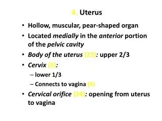
UTERUS UTERUS LOCATED • Inside the pelvis • Dorsal to the urinary bladder • Ventral to rectum
Part of uterus Body of uterus • Fundus • Cavity of uterus Function • accept a fertilized ovum which passes through the utero-tubal junction from the fallopian tube
Neck of uterus ( cervix uteri) • External orifice of the uterus • Canal of the cervix • Internal orifice of the uterus Function • to allow sperm and menstrual fluid to move through.
Layer of uterus Uterus consists of 4 layers : • Endometrium- innermost glandular layer • Myometrium- smooth muscle located between the endometrium and perimetrium. • Perimetrium-The peritoneum covering of the fundus and ventral and dorsal aspects of the uterus. • Parametrium-The loose connective tissue around the uterus.
There are four main forms of uterus in mammals. They are: Duplex Bipartite Bicornuate Simplex
Duplex uterus • Two wholly separate uteri, with one fallopian tube each which opens into the vagina. Found in marsupials, rodents, and lagomorphs opossums Tasmanian devils
Absent (or limited) fusion of the Mullerianducts leads to the presence of two uteri. The urogenital sinus (US) is connected to the female reproductive tract • In monotremes rather than nurturing the embryo, the uterus secretes the shell around the egg. It is essentially identical with the shell glandof birds and reptiles, with which the uterus is homologous.
Mullerian duct fusion is physically blocked by the presence of the ureters, which leads to the formation of three vaginae • The duplex uterus shown here has a pair of cervices.
Bipartite uterus • The two uteri are separate for most of their length, but share a single cervix. Found in ruminants (deer) and cats. • Mullerianfusion in the uterine region does not occur, or is limited, which leads to the formation of a pair of uterine horns that can support the development of many fetuses.
Bicornuate uterus • The upper parts of the uterus remain separate, but the lower parts are fused into a single structure. • A larger portion of the uterus forms the uterine body. Prosimian primate
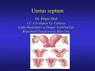
Simplex uterus • The entire uterus is fused into a single organ. Found in higher primates. • some individual females (including humans) may have a bicornuate uterus, a uterine malformation where the two parts of the uterus fail to fuse completely during fetal development.
Essential in sexual response by directing blood flow to the pelvis and to the external genitalia • To accept a fertilized ovum which passes through the utero-tubal junction from the fallopian tube. It implants into the endometrium, and derives nourishment from blood vessels which develop exclusively for this purpose. • The embryo attaches to a wall of the uterus, creates a placenta, and develops into a fetus (gestates) until childbirth.
histology of uterus Uterus has 3 layers: Perimetrium (outer) – tunica serosa Myometrium – tunica muscularis Endometrium – tunica mucosa
perimetrium • tunica serosa • It has the composition of loose connective tissue • contains a large number of lymphatic vessels
myometrium • tunica muscularis of the uterus. • composed of a thick inner circular layer and a thinner outer longitudinal layer of smooth muscle. • The region in between the two layers of smooth muscle contains large blood vessels. • Stratum vasculare- layer of large blood vessels located between the inner and outer layers of smooth muscle of the myometrium. • In the sow the stratum vasculare is indistinct and in the cow it may be located in the outer half of the circular muscle layer.
endometrium • comprises the tunica mucosa and the tunica submucosa • tunica mucosa - lamina epithelialis is usually simple columnar except in the sow and ruminants (pseudostratified columnar) • The lamina propria- loose connective tissue full of neutrophils and lymphocytes. • It blends with the underlying tunica submucosa since there is no lamina muscularismucosae in the entire female reproductive tract. • Uterine glands are simple or branched tubular glands located in the lamina propria-tunica submucosa. • Some regions of the endometrium in ruminants are void of glands and are highly vascular. It is in these regions, called caruncles, that contacts between the uterus and the extraembryonic membranes are made. • These glands provide nourishment for the early stages of embryonic growth, before the placenta is established. Structurally - simple tubular in late pregnancy - tortuous and coiled in shape.
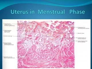
Key1. Lumen 2. Endometrium lined by cuboidal epithelium3. Lamina propria4. Endometrial glands 5. Myometrium6. Serosa H&E, 40x
Key1. Lumen 2. Endometrium3. Endometrial glands 4. Myometrium, circular section5. Stratum vasculare6. Myometrium
FALLOPIAN TUBES WANNURSYAMIMI BTE WAN ZAHARI- D11BO46 HERLINA BINTI MOHD RAPI – D11A010 NIK NUR AFINA BTE NIK ALWI –D11A021
Fallopian Tubes (or Oviducts) • pair of small tubes leading from the ovaries to the horns of the uterus • Millions of tiny hair-like cilia line the fimbria and interior of the fallopian tubes • Size - long(5 - 6 inches) - diameter (0.2-0.6 inches) • Fertilization occurs in the oviduct. • The journey through the Fallopian tube takes about 7 days. • Because the oocyte is fertile for only 24 to 48 hours • The cilia beat in waves hundreds of times a second catching the egg at ovulation and moving it through the tube to the uterine cavity
Fimbrial segment -faces the ovary • Infundibular segment - funnel shaped segment behind the fimbria • Ampullary segment -wide middle segment • Isthmic segment - narrow muscular segment near the uterus • Interstitial segment - passes through the uterine muscle into the uterine cavity
OVIDUCT FALLOPIAN TUBE Reproductive system of avian Reproductive system of mare
Roles each parts in fallopian tubes • The infundibulum catches and channels the released eggs. • The ampulla is the site where fertilization occur. • The endings of the fimbriae extend over the ovary; they contract close to the ovary’s surface during ovulation in order to guide the free egg. • The isthmus connects the ampulla and infundibulumto the uterus.
GENERAL FUNCTIONS OF FALLOPIAN TUBE WHAT IS UTERUS? Provide a place for fertilization between egg and sperm. Site for transportation of the egg from the ovary. mucous membrane lining the fallopian tube gives off secretions that help to transport the sperm and the egg and to keep them alive.
Histology of Fallopian Tube ( tuba uterina , oviduct) • The uterine tubes are long flexuous musculomembranous tubes. • They consist of: • Infundibulum: an expanded cranial end which is close to the ovary; • Ampulla: a middle segment • Isthmus: a caudal narrow part that opens into the ipsilateral horn of the uterus. • The epithelium is simple or pseudostratified columnar with secretory and ciliated cells. • The lamina propria has longitudinal folds giving it a glandular appearance. • In some breeds of sheep (blackface and crosses), pigment cells are present. • The muscularis is mainly a layer of circular smooth muscle, thickening at the junction with the uterine horn, with an outer layer of longitudinal muscle. • The serosa is loose vascular connective tissue with prominent blood vessels
Serosa Folds Muscularis
Uterine tube in sheep Transverse section ; Stain: Hematoxylin-eosin ; x62.5
Lumen : lined by pseudostratified epithelium Muscularis : most part circular smooth muscle fibres Lamina propria Serosa : loose vascular connective tissue with prominent blood vessels
Uterine Tube of Cow Transverse section ; Stain : Hematoxylin-eosin ; x125
Deep mucosal folds are cut in transverse section giving the appearance of mucosal glands. Pseudostratified columnar with Ciliated and nonciliated cells.
Oviduct – AmpullaTubaeUterinae Stain : iron hematoxylin-eosin ; magnification : x10
Oviduct musculature Oviduct musculature Mesosalpinx Tunica serosa artery Vein Mucosa fold (plica)
Oviduct – Isthmus TubaeUterinae Stain: alum hematoxylin-eosin ; magnification : x40
Smooth Musculature Mucosa plicae Artery Subserosal tissue Tunica Serosa Smooth musculature
Oviduct- Uterina part of the oviduct Stain : hematoxylin-eosin; magnification: x25