Download
1 / 17
190 likes | 629 Vues
Measurement & Microscopes. Size relative Prefix Meaning to a meter Length Kilo- thousand 10 3 kilometer (km) Centi- hundredth 10 -2 centimeter (cm) Milli- thousandth 10 -3 millimeter (mm) Micro- millionth 10 -6 micrometer (µm) Nano- billionth 10 -9 nanometer (nm).

E N D
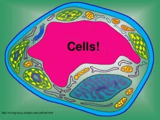
Size relative Prefix Meaning to a meter Length Kilo- thousand 103 kilometer (km) Centi- hundredth 10-2 centimeter (cm) Milli- thousandth 10-3 millimeter (mm) Micro- millionth 10-6 micrometer (µm) Nano- billionth 10-9 nanometer (nm) Sizes of Things 4 mm 2 mm 200 µm • With the unaided eye, you can see down to about 200 µm (that’s 0.2 mm). • With a quality light microscope, you can see something as small as 200 nm clearly (that’s 0.0002 mm). • With an electron microscope, you can visualize something as small as 0.2 nm clearly (that’s 0.0000002 mm or otherwise stated, 2x10⁻⁷ mm). 200 nm Check out: http://www.cellsalive.com/howbig.htm
Sizes of Things & Photomicrography Check out: • How Big? • http://www.cellsalive.com/howbig.htm • Scale of the Universe • http://htwins.net/scale2/ • Olympus Bioscapes Competition winners • http://www.olympusbioscapes.com/gallery/2012/ • Nikon Small World Competition (photo & video) • http://www.nikonsmallworld.com/
Microscopes: Tools of Biologists Different microscopes have different advantages and disadvantages. Biologists use different microscopes depending upon what they want to look at / observe. • ________________________: also called a stereo microscope • 2 eyepieces • 2 lenses, __________ and ___________ (in the eyepiece) • 2 lamps, one above and one below the specimen • Advantages: • Specimen can be alive • No special preparation of specimen needed • Easy to use! • Relatively inexpensive • Disadvantages: • Low magnifying power (up to 60x usually) Dissecting Microscope objective ocular lens
Compound Light Microscope ocular lens objective water • ______________________________ • 2 lenses, __________ and ___________ (aka eyepiece) • On student ‘scope, usually have 10x ocular lens & several objective lenses: 2x (scanning), 10x (low power), & 40x (high power) • Sample often placed in _______ on a slide • Some specimens are stained or “fixed” with dyes -- this enhances contrast & helps preserve specimen (but kills it too) • Light passes _________ sample and is “bent” by lenses to produce a magnified image • Advantages: • Specimen can be alive • Easy specimen preparation • Relatively easy to use • Fairly inexpensive for a basic ‘scope • Can magnify up to _______ clearly • Disadvantages: • Cannot see anything smaller than ______________ (note: a small cell is about 10 µm across) through 1000x 0.2 µm (200 nm)
Modern Optical Microscopes with Digital Camera attachments http://www.microscopyu.com/museum/e600pol.html http://www.microscopyu.com/museum/smz1500.html
Transmission Electron Microscopy http://www.fei.com/products/tem/tecnai/ http://en.wikipedia.org/wiki/Transmission_electron_microscopy
In TEM, the specimen must be thin enough to allow electrons to travel through the tissue. You can cut very thin slices of your specimen either by fixing it in plastic or freezing it. The specimen is cut into thin sections by a diamond knife in an instrument called ultramicrotome. Each section is only 50-100 nm thick. A thin section of your sample is placed on a copper grid and stained with a heavy metal, like uranium & lead, which scatters electrons well and improves the contrast in the microscope. The slice of tissue can now be studied under the electron beam. Alternately, a solution of isolated material (can be a solution with bacteria or a solution with isolated molecules) is spread on a support grid coated with plastic. A solution of heavy metal salt is added. The metal salt solution does not bind to the material but forms a "shadow" around it on the grid. The specimen will appear as a negative picture when viewing it in the TEM. TEM Specimen Preparation An ultra-microtome slices samples as fine as 70 nm thin so they can be viewed in a transmission EM. http://www.eicn.ucla.edu/UTCultramicrotome http://www.nobelprize.org/educational/physics/microscopes/tem/preparation.html http://en.wikipedia.org/wiki/Transmission_electron_microscopy
Scanning Electron Microscopy http://www.nist.gov/cnst/nanofab/toolset-metrology.cfm http://en.wikipedia.org/wiki/Scanning_electron_microscope
• Electron Microscopes electrons electromagnets • Sends beam of _________ over or through specimen • This beam is bent by large _______________ to produce a magnified image There are two main types of electron microscopes: • _____________ Electron Microscope (TEM) • Beam of electrons passes ________ an ultra-thin section of a specimen and onto a fluorescent screen or photographic film • Images produced are ________________ of specimens • __________ Electron Microscope (SEM) • Beam of electrons passes over the _______of a specimen, electrons bounce off at different angles & are “read” to produce image • Images are ____ pictures of the ________ of samples (no inside views) Transmission through cross-sections Scanning surface 3D surface
TEM ____ Micrographs Pancreatic cell Golgi body http://en.wikipedia.org/wiki/Transmission_electron_microscopy Bacillus subtilis bacterium http://www.histology-world.com/photoalbum/displayimage.php?album=55&pid=509#top_display_media Herpes virus Nucleus surrounded by rER Mitochondria (in lung cells) http://www.histology-world.com/photoalbum/thumbnails.php?album=55&page=2
http://micro.magnet.fsu.edu/primer/java/electronmicroscopy/magnify1/index.htmlhttp://micro.magnet.fsu.edu/primer/java/electronmicroscopy/magnify1/index.html SEM ____ Micrographs Can you guess what this is? Hair shaft Ebola Viruses (one w/ false color) The fossilized shell of a microscopic ocean animal, magnified 392 times its actual size. The ancient creature, called Radiolarian, lived in the waters off Antarctica and is now used to study such things as climate and ocean circulation. http://bepast.org/dataman.pl?c=lib&dir=docs/photos/ebola/
What is this? http://www.aecom.yu.edu/aif/gallery/sem/sem.htm
White blood cell (B-cell) with viruses budding out of it. http://www.aecom.yu.edu/aif/gallery/sem/sem.htm
TEM (above) and SEM (below) of viruses budding from a mammalian cell http://www.aecom.yu.edu/aif/gallery/sem/sem.htm
• Electron Microscopes viruses sub-cellular organelles • Advantages: • Can be used to view smaller things like ________ and _____________________ that cannot be seen with light ‘scopes • Can magnify over 100,000x and allows you to see objects as small as _______ • Disadvantages: • Preparation of specimen can be _________ Ex) • Cannot view living organisms • Much more complex & difficult to use/operate than light ‘scope • Extremely expensive to buy and maintain • Only produces ______________ images (any color electron micrographs have false color added afterwards by computer) 0.2 nm difficult Freezing or embedding in resin, slicing ultra-thin sections black & white
Virtual Electron Microscopy http://micro.magnet.fsu.edu/primer/java/electronmicroscopy/magnify1/index.html