Download
1 / 45
500 likes | 1.21k Vues
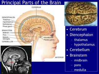
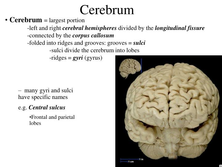
Cerebrum. Cerebrum = largest portion -left and right cerebral hemispheres divided by the longitudinal fissure -connected by the corpus callosum -folded into ridges and grooves: grooves = sulci -sulci divide the cerebrum into lobes -ridges = gyri (gyrus).

E N D
Cerebrum • Cerebrum= largest portion • -left and right cerebralhemispheres divided by the longitudinal fissure • -connected by the corpus callosum • -folded into ridges and grooves: grooves = sulci • -sulci divide the cerebrum into lobes • -ridges = gyri(gyrus) • many gyri and sulci have specific names • e.g. Central sulcus • Frontal and parietal lobes
Cerebrum • -cerebrum is comprised of white and • gray matter: • 1. white matter - neurons with • long, myelinated axons • -organized into tracts • -three categories of tracts • commisural – join areas between hemispheres • e.g. corpus callosum • b. association – joins areas within • a hemisphere • c. projection – joins cerebrum to brain stem
Protection: CSF • brain contains fluid-filled chambers = Ventricles • Chambers in central passageway of the brain • 2 lateral ventricles, 1 third ventricle, 1 fourth ventricle • connects to the central canal which runs into the spinal canal • These chambers contain cerebrospinal fluid
I III II V VI VII IX VIII X
Cervical • and lumbar • enlargements
Dorsal Ramus Ventral Ramus Spinal nerve Ventral Root Dorsal Root Dorsal Root ganglion
Dorsal Ramus Dorsal Root Ventral Ramus Ventral Root Rami Communicantes
Right Atrium • Receives blood from 3 sources • superior vena cava, inferior vena cava and coronary sinus • Interatrial septum partitions the atria • Fossa ovalis is a remnant of the fetal foramen ovale • Tricuspid valve • Blood flows through into right ventricle • has three cusps composed of dense CT covered by endocardium Right atrium Interatrial septum Tricuspid AV valve
Papillary muscle Chordae tendinae
SA node Bundle of His AV node Bundle branches Purkinje fibers
Left Common Carotid Brachiocephalic Trunk Left Subclavian Superior Vena Cava Aortic Arch Ascending Aorta Pericardium
Left Common Carotid Left Subclavian Aortic Arch Parietal Pricardium Diaphragm
Right Conus artery Left coronary artery Left Marginal Artery & vein Right coronary artery Anterior Interventricular Small cardiac vein Great Cardiac Vein Right Marginal artery Anterior Interventricular Green dots on veins
Great Cardiac Vein Circumflex artery Posterior Interventricular Artery (right & left) Coronary Sinus Green dots on veins
vertebral thyrocervical suprascapular thoracoacromial Common Carotid subscapular circumflex humeral deep radial brachial radial collateral ulnar collateral brachial radial ulnar
Radial collateral Ulnar collateral brachial Common interosseous radial ulnar interosseous Deep palmar arch Superficial palmar arch Digital arteries
posterior auricular superficial temporal maxillary occipital internal carotid external carotid carotid sinus facial lingual superior thyroid
Inferior Vena Cava Celiac Superior Mesenteric Renal Gonadal Inferior mesenteric Common Iliac
Left Gastric Splenic Hepatic Proper Splenic Vein Common Hepatic Celiac trunk Inferior Mesenteric
Left Colic Artery Sigmoid Superior Rectal
Common iliac External iliac Internal iliac Ascending br Of Lateral circumflex Lateral circumflex Obturator Deep femoral Medial circumflex Descending br Of Lateral circumflex Femoral
Deep Femoral Femoral Genicular Descending br Of Lateral circumflex Genicular Arteries of the Knee Anterior Tibial
Eustacian tube With tubal tonsil
Parotid Sublingual Submandibular
Thoracic Aorta Esophagus Diaphragm Liver
FUNDUS LESSER CURVATURE BODY GREATER CURVATURE PYLORIC REGION PANCREAS DUODENUM
Lesser Omentum Liver Stomach Gallbladder Descending Colon Transverse Colon Ascending Colon Mesentery of Small Intestine Small Intestine
Right & Left Hepatic Ducts Common Hepatic Duct Pancreatic Duct Gallbladder Cystic Duct Ampulla of Vater Common Bile Duct
Hepatic Portal Vein Splenic Vein Common Bile Duct Superior Mesenteric Vein Inferior Mesenteric Vein
kidney URETER URETER BLADDER
Renal Papilla Renal Pyramid Minor Calyx Major Calyx Renal Cortex Renal Pelvis Renal Medulla URETER
Bladder Rectum Prostate Corpus spongiosum Prostatic Urethra Membranous Urethra Corpus cavernosum Spongy/ Penile Urethra Glans Penis Testes External Urethral Orifice
Corpus cavernosum Corpus spongiosum Spongy/ Penile Urethra Vas Deferens Epididymus Spermatic Cord Testes
pampiniform plexus Vas deferens testicular artery Epididymus Seminiferous Tubules Tunica Albuginea Tunica Vaginalis -testis: develop internally near the kidneys and descend through the inguinal canal during the latter half of the seventh month gestation -covered by several protection membranes 1. tunica vaginalis – serous membrane derived from the peritoneum, forms during the descent of the testes -injury to the testes can cause an accumulation of fluid within the membrane = hydrocele -allows for easier movement of the testes within the scrotum 2. tunica albuginea – internal to the TV -extends inward to divide the testes into lobules (200-300) -each lobule contains 1 to 3 coiled seminiferous tubules for sperm production
Round ligament Broad ligament Fundus Fundus Ovary Bladder Fornix Body Fimbrae of oviduct Fallopian Tube (oviduct) Cervix Bladder Rectum Vaginal canal Urethra External urethral orifice Labia minora Vaginal orifice Labia majora