Download
1 / 36
630 likes | 1.01k Vues
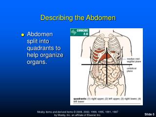
The Abdomen. Stuart M Bunt. Functional Anatomy 212. Overview. Embryology Revision. Foregut, Midgut and Hindgut suspended by the dorsal mesentary, initially straight Ventral mesentary connects stomach and ant. abd. wall, rest of gut free anteriorly

E N D
The Abdomen Stuart M Bunt Functional Anatomy 212
Embryology Revision • Foregut, Midgut and Hindgut suspended by the dorsal mesentary, initially straight • Ventral mesentary connects stomach and ant. abd. wall, rest of gut free anteriorly • Mesentary supplies blood and nerves to gut between layers of peritoneum • Complex adult layout due to 270o rotation
Blood Supply to Abdominal Organs • Foregut • Celiac trunk • Midgut • Superior mesenteric artery • Hindgut • Inferior mesenteric artery • Rectum • Internal iliac artery (pudendal and rectal arteries)
Stomach • Variable size and shape, distensible • J shaped related to body form • Lesser and greater curvature • gastroesophageal junction • fundus,cardiac part, body, pyloric part • pyloric antrum and sphincter • rugae and gastric pits
Venous system • Portal Vein • Splenic vein • inferior mesenteric vein • Superior mesenteric vein • Gastric veins • Hepatic Veins Inf. Vena Cava
Stomach rotates and distends Front Dorsal Mesentary Ventral Mesentary Back Splenic tissue Omentum Epiploic Foramen
The Peritoneal cavity is divided in two • Rotation of stomach forms the greater omentum (allows stomach distension and infection control) • Omental bursa or Lesser sac is inside omentum (a potential space) • Lesser omentum runs from stomach to liver (note free lower border above epiploic foramen contains portal vein, hepatic artery and bile duct • Falciform ligament runs from liver to ant abd. wall
Mesenteries are important:- • Paracolic gutters channel fuid • Stop herniation due to bipedal posture • Supply blood/nerves • Sensitive to stretch • Contain infection • Useful in surgery
On return some gut fuses with posterior wall Diaphragm 1 2 Duo. Asc. Colon Desc. Colon 3 1.lienorenal lig. 2.trans. mesocolon 3.mesentary proper 4.mes. of sig. colon 4 Rectum
Oesophagus • 10 inches from pharynx to stomach • narrow • at cricoid cartilage • where left bronchus crosses • oesophageal hiatus in diaphragm • mucous membrane folded (normally collapsed) • stratified squamous epithelium • striated above smooth below • trachea on right, • lower aorta on left • medial to L. lung, behind left atrium
Duodenum • first 12 inches of gut • four parts form C shape • duodenal cap • radiologically identified, ulcers form here • mobile • descending part • pancreatic and bile ducts • horizontal part • crosses psoas, IVC and aorta • crossed by mesentery, sup mesen. art. • ascending part
Jejunum • 2/5ths of small intestine • gradual transition to ileum • many small villi • increasing numbers of lymph nodules • no submucosal glands • lacteals in each villus • columnar epithelium
Ileum • distal 3/5ths of intestine • narrower, thinner, less vascular, slower, • more fat and arterial arcades in mesentery than jejunum. • Peyer’s patches of lymphoid tissue
Colon • ascending colon retroperitoneal • right colic or hepatic flexure • transverse colon (mesocolon) • droops towards pelvis? • left colic or splenic flexure • descending colon retroperitoneal • pelvic or sigmoid colon S shaped
The Liver • Largest Gland (one of largest organs) • Right upper abdomen under diaphragm • Grows as outgrowth of gut plus mesoderm • Diaphragmatic surface • Visceral surface down and left • related to stomach, duodenum, r. kidney, r. colonic flexure • bears gall bladder
Biliary System • R and L Hepatic ducts • Common hepatic duct • Joined by cystic duct (to gall bladder) • Forms bile duct (common bile duct) • Gall Bladder • body and fundus, salts and water absorbed • store for bile, released in response to cholecystokinin
Pancreas • Head in concavity of duodenum • body across vertebrae • tail reaches the spleen • pancreatic duct (+ accessory?) • ampulla • duodenal papilla
The Spleen • Lies in left hypochondriac region between gastric fundus and diaphragm at level of 9th-10th rib (not normally palpable) • Soft, friable, highly vascular, dark purple • Diaphragmatic surface • convex and smooth facing diaphragm • Visceral surface • gastric, renal, pancreatic and colic impressions
The Spleen (2) • Hilum of spleen long fissure through which vessels and nerves pass • Suspended from stomach by gastrolienal ligament (contains short gastric and left gastro-epiploic branches of spenic artery) • Suspended from posterior abdominal wall by lienorenal ligament • Covered by adherent peritoneum
Kidneys In fat capsule Suprarenal glands superiorly Direct Arterial and venous supply
Kidney Internal Structure • Renal pyramids between renal columns • Renal Cortex • Renal papillae drain into minor calix • Major calix join to form renal pelvis • Ureter as outlet
Kidneys External View • Artery - Vein - Ureter