Download
1 / 34
340 likes | 468 Vues
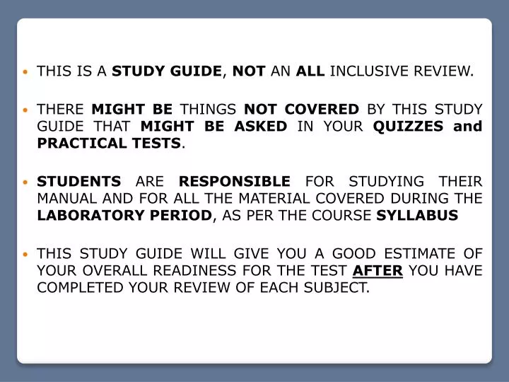
THIS IS A STUDY GUIDE , NOT AN ALL INCLUSIVE REVIEW. THERE MIGHT BE THINGS NOT COVERED BY THIS STUDY GUIDE THAT MIGHT BE ASKED IN YOUR Q UIZZES and PRACTICAL TESTS .

E N D
THIS IS A STUDY GUIDE, NOT AN ALL INCLUSIVE REVIEW. • THERE MIGHT BE THINGS NOT COVERED BY THIS STUDY GUIDE THAT MIGHT BEASKED IN YOUR QUIZZES and PRACTICAL TESTS. • STUDENTS ARE RESPONSIBLE FOR STUDYING THEIR MANUAL AND FOR ALL THE MATERIAL COVERED DURING THE LABORATORY PERIOD, AS PER THE COURSE SYLLABUS • THIS STUDY GUIDE WILL GIVE YOU A GOOD ESTIMATE OF YOUR OVERALL READINESS FOR THE TEST AFTERYOU HAVE COMPLETED YOUR REVIEW OF EACH SUBJECT.
Lab # 9 Nervous System - Neural Tissue - Spinal Cord - Spinal Nerves
NEURON STRUCTURE -In the CNS -In the PNS Histology of the Nervous Tissue 1- Neurons Glial cell (astrocyte) 2- Neuroglia or glial cells Neurons The glial cells are supporting cells, which are associated to the neurons and provide a supportive scaffolding for neurons
NEURON STRUCTURE Free ribosomes Axon R.E.R. Axon hillock Telodendria Axon terminal or synaptic terminal Dendrites The Structure of Neurons Nissl bodies It carries the nerve impulses away form the soma. Protein synthesis Neurofibrils Nucleus It is the trigger zone for the nerve impulse. Cell body or Soma Perikaryon Telodendria They provide internal support for the extensions and are responsible for intracellular transport. Axon Axon terminal , synaptic terminal, or synaptic knob Dendrite Synapses with another neuron and releases the neurotransmitters. They receive the nerve impulse.
Neurilemma Schwann cell Nucleus of Schwann cell Axoplasm Axolemma Myelin sheath Nodes of Ranvier It is the plasma membrane of the Schwann cells. It is the plasma membrane of the axon. It is the cytosol of the axon. They produce the myelin sheath. It electrically insulates the axon and enables saltatory conduction. It is where the depolarization of the membrane occurs during saltatory conduction.
Synapse from neuron to neuron. Synapse from neuron to effector cell. It is a junction that mediates information transfer from one neuron to the next or from a neuron to an effector cell (ex: muscle cell, gland). Synapse: Neuromuscular junction
The Synapse The operation of the nervous system depends on the flow of information through chains of neurons functionally connected by synapses. It conducts impulses towards the synapse. It conducts impulses away from the synapse. Presynaptic neuron Postsynaptic neuron
Structural Classification of the Neurons Locations: Brain and special sense organs. Locations: Special sen- se organs (retina, olfactory epithelium). Functions: Poorly understood. Functions: They relay information about sight, smell or hearing from receptors cells to other neurons. Locations: Dorsal root ganglia of spinal cord. Locations: Anterior gray horn of spinal cord, primary motor cortex of the cerebrum. Functions: Most sensory neurons of the PNS. Functions: Motor neurons that control skeletal muscles.
1- Sensory or afferent neurons 2- Motor or efferent neurons 3- Association neurons or Interneurons Functional Classification of the Neurons They carry information towards the Central Nervous System. They carry information away from the Central Nervous System. They carry impulses between sensory and motor neurons located at CNS.
Spinal Cord and Spinal Nerves
Gray matter Cell bodies of neurons and glia (no myelin). It forms nuclei in the CNS White matter Myelinated fibers. It forms tracts and nerves in the Peripheral Nervous System White matter Gray matter
The Anatomical Divisions of the Nervous System • Brain • Central Nervous System (CNS) • Peripheral Nervous System (CNS) • Spinal cord • It consists of the brain and spinal cord enclosed by cranium and vertebral column. • Nerves • Ganglia • It is all the nervous system except the brain and spinal cord. • It consists of nerves and ganglia. It is responsible for integrating, processing and coordinating sensory data and motor commands. It deliveries sensory information to the CNS and carries motor commands to peripheral tissues and system. Nerve: It is a bundle of nerve fibers (axons) wrapped in fibrous connective tissue. It is a bundle of nerve fibers (axons) in the CNS (white matter). Tract: • It is a concentration of neuron cell bodies in the CNS (gray mater). Nucleus: • It is a knot-like swelling in a nerve where neuron cell bodies are concentrated. Ganglion:
Foramen magnum Cervical enlargement Posterior median sulcus Lumbar enlargement L1 – L2 Cauda equina Conus medullaris Filum terminale Gross Anatomy of Spinal Cord (Fibrous tissue that avoids up and down movements of spinal cord)
Dura matter Arachnoid Pia matter Arachnoid Pia matter Dura matter Epidural space Subarachnoid space Denticulate ligament Subdural space Spinal meninges: Dura matter, arachnoid matter, pia matter (It is filled with CSF) (Epidural anesthesia) (Prevent lateral movement)
Lumbar puncture (spinal tap) is the most common means of collecting a specimen of cerebral spinal fluid. The spinal needle is inserted, usually between the 3rd and 4th lumbar vertebrae. Once the needle is properly positioned in the subarachnoidspace, pressures can be measured and fluid can be collected for testing.
Dorsal or posterior root Dorsal or posterior root ganglion Posterior gray commissure Posterior gray horn Somatic sensory Lateral gray horn Anterior gray horn Somatic motor Anterior gray commissure Spinal nerve Ventral or anterior root Anterior median fissure GRAY MATTER ORGANIZATION POSTERIOR Posterior median sulcus Visceral sensory Visceral motor ANTERIOR
Somato-sensory neurons Cerebral cortex Cerebral cortex Visceral-sensory neurons Thalamus Hypothalamus Somato-motor neurons Visceral-motor neurons SS VS VM SM Somatic sensory fiber Somatic motor fiber Visceral sensory fiber Visceral motor fiber (Autonomic Nervous System)
Central canal Anterior white commissure WHITE MATTER ORGANIZATION POSTERIOR (Contains CSF) Posterior white column Lateral white column Anterior white column ANTERIOR