Download

1 / 41
410 likes | 611 Vues
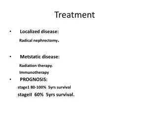
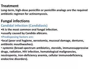
Treatment. Treatment Goals include: Stabilize patient Treat abdominal trauma and possibly damaged underlying structures Liver Diaphragm Kidney Small Intestines Large Intestines. Treat R Leg gunshot wound Prevention of complications of injury and surgery Treat fracture and Pain

E N D
Treatment • Treatment Goals include: • Stabilize patient • Treat abdominal trauma and possibly damaged underlying structures • Liver • Diaphragm • Kidney • Small Intestines • Large Intestines
Treat R Leg gunshot wound • Prevention of complications of injury and surgery • Treat fracture and Pain • Rehabilitation • Treatment of Ulcers • Treatment of the following conditions if established • Hemothorax • Post-Traumatic Stress Disorder
Initial Assessment • ABCDE • A : Airway and C-Spine Control • B : Breathing • C: Circulation • Assess vital signs • Control bleeding • D: Disability • Assess GCS, pulses, sensory and motor functions • E: Exposure and Environment • Undress, Keep warm, Check parts often missed
Management of Abdominal Trauma • Initial dressing and Antibiotics • sterile cover should be given and left in place until wound can be seen in the Operating Room • Benzylpenicillin • mainstay of first line treatment • especially for soft tissue injuries • Clostridia and Beta-haemolytic streptococci caused major fatal infective complications in war wounds
Management of Abdominal Trauma • Penetrating Abdominal Trauma • Surgical management has been the general standard of care for penetrating abdominal injuries • in the absence of hemodynamic instability or signs of hollow viscus perforation, the majority of abdominal stab wounds and many gunshot wounds can initially be managed nonoperatively
Management of penetrating abdominal trauma. CT = computed tomography; DPL = diagnostic peritoneal lavage; RBC = red blood cells. * Image from http://img.medscape.com/pi/emed/ckb/trauma/432648-1364845-433554-433665.jpg
Management of Abdominal Trauma • General Considerations in Penetrating Abdominal Trauma • Little pre-op evaluation required for GSWs that penetrate the peritoneal cavity • Chance of internal injury over 90% • Laparotomy is mandatory • All abdominal explorations in adults are performed using a long midline incision • incision made with a scalpel (faster) rather than electrosurgical unit
Trauma Laparotomy: Essentials • Initial Control of Bleeding • accomplished with 4 quadrant packing using laparotomy pads (placed above liver, spleen and in both sides of pelvis) • once anesthesia has been given time to catch up with fluid resuscitation, the packs are removed one quadrant at a time, starting away from sites of apparent bleeding • Identification of Injuries • each solid organ and the entire bowel are inspected • Control of Contamination • with clamps, staples, or suture closures • Reconstruction • depending on character of defect(s), resection may be necessary • if patient is stable enough to continue the operation, reconstruction may then be performed
Management of Abdominal Trauma • Liver • Control bleeding and bile leak (if present) • Control infection (esp. in patients with associated visceral injury) • Remove devitalized tissue
Management of Abdominal Trauma • Operative management of liver injuries can involve many techniques: • simple packing or wrapping • thrombin, fibrin sealant, collagen/gel preparations, electrocautery, argon beam and radiofrequency coagulation, omental packing, or even intrahepatic balloon tamponade as in the case of through-and-through injuries. • local hemostasis • resectional debridement • finger fracture, cautery, sutures, clips, or stapler device
Management of Abdominal Trauma • Diaphragm • Penetrating injuries to the diaphragm are graded as follows: (I) contusion (II) laceration, <2 cm (III) laceration, 2-10 cm (IV) laceration, >10 cm (V) total tissue loss, >25 cm2. • Lower grade injuries may be repaired either via laparotomy or with laparoscopic or thoracoscopic techniques.
Essential components of repair include: • airtight closure with nonabsorbable suture • liberal saline lavage of the hemithorax if there has been a concomitant bowel injury with soilage of the field. • chest tube is often placed for drainage • large defects may require placement of a prosthetic patch
Management of Abdominal Trauma • Kidney • Injuries to the kidney are also graded according to severity, as follows: • (I) contusion • (II) lacerations, <1 cm • (III) lacerations, >1 cm • (IV) lacerations to the collecting system • (V) vascular avulsion
kidney is salvaged with renography, using: • pledgeted sutures and wrapping • capsular reapproximation (if at all possible) • If nephrectomy is deemed necessary because of the severity of injury or instability of the patient the contralateral kidney should be checked using IV pyelogram
Management of Abdominal Trauma • Duodenum • Injuries to the duodenum are graded as follows: • (I) hematoma; • (II) partial thickness laceration • (III) laceration disrupting <50% circumference of D1, D3, D4, or 50-75% circumference of D2 • (IV) laceration disrupting 50-100% circumference of D1, D3, D4, or >75% circumference of D2, or involving the ampulla or distal common bile duct • (V) massive disruption of the duodenopancreatic complex or devascularization of the duodenum.
The Kocher maneuver used to mobilize the duodenum, along with the pancreatic head and distal common bile duct, to explore penetrating injuries • Primary repair of injury is the goal, with protection of the repair using closed-suction drainage
Diversion procedures are often used for protection • Duodenal diverticularization • Pyloric exclusion • Grade V injuries require pancreaticoduodenectomy • done as a staged procedure in the unstable trauma patient
Treatment of Abdominal Trauma • Small Bowel • Control of contamination is of high priority with penetrating injuries to the small bowel. • Clamps or staples may be used for temporary control as the entire length of the small bowel is examined
Management of Abdominal Trauma • Colon • management of colonic injuries depends on the: • extent of the defect • the amount of contamination • stability of the patient • Primary repair may be considered if the patient is hemodynamically stable and if the injury is fairly small with minimal fecal contamination.
a diverting colostomy should be performed if: • patient has multiple injuries • patient has required significant blood product resuscitation • if the patient is acidotic, hypothermic, and coagulopathic • and/or if there is a large defect (>50% of the circumference) and considerable fecal spillage
Management of Abdominal Trauma • Post-operative details • Patients should be monitored closely in the surgical intensive care unit after trauma laparotomy • Patient may still be intubated and could require ventilatory support • Continue management: • fluids and blood product resuscitation • Replace electrolytes • Monitor drain outputs
Management of R Leg gunshot wound • Wound Surgery • Incision and irrigation. An incision should pass through the skin wound, trimming only its grossly damaged edges, and continue in the axis of the limb, crossing flexor creases obliquely. • Damaged subcutaneous fat and shredded fascia are removed. The deep fascia is incised for the length of the incision or beyond it, to allow exploration and relieve pressure within the wound and associated compartments.
Management of R Leg gunshot wound • Irrigation with copious volumes of saline is used to reduce the number of bacteria, and pulsating high-pressure irrigation may be even more effective. • Excision. Muscle is assessed for color, consistency, contractility and capacity for bleeding. • Dressing and closure. Dressing the open wound with fluffed-out gauze allows drainage with no need for a surgical drain. • Suturing is appropriate only if all tissues appear healthy and the edges of the skin and deeper tissues can be approximated without undue tension.
Management of R Leg gunshot wound • Splintage • Even when there is no fracture, the injured limb needs support and stabilization by a plaster cast or backslab, to protect the soft tissues.
Prevention of complications associated with the injury and surgery • Antibiotics • Coverage for Staphylococcus sp. and anaerobes • Tetanus prophylaxis • Tetanus Toxoid • Prophylaxis for venous thromboembolism • Heparin
Fracture and Pain Management • Fractures • Fixation • Calcium Supplementation recommended daily intake: 1000-1200mg • Vitamin D supplementation • Physical activity • Pain • Opioids
Rehabilitation • Range of Motion exercises • Strengthening Exercises • Isometrics • Weight training • Pulmonary Rehabilitation • diaphragmatic breathing exercises • ventilatory muscle training • pacing – performance of an activity within the limits or boundaries of that patient’s breathing capacity • Bed mobility/transfers training • Gait training • ADL training (also with an occupational therapist)
Rehabilitation • Modalities: • Hydrotherapy • Facilitate patient in doing lower extremity exercises • Also, for mechanical debridement of wound • Compression therapy: intermittent pneumatic compression • Compression assists with venous return and stimulates the release of cellular factors that facilitate wound healing • Electrical stimulation: • Neuromuscular Electrical stimulation/Functional Electrical stimulation • to facilitate neuromuscular recovery/healing • Radiant Heat • Infrared radiation increase local wound and skin temperatures facilitating higher metabolic rates and improving circulatory activity of the wound
Management of Ulcers Medical CareThe treatment of ulcerated lesions varies depending upon size, duration, and location.✔With ulcerations induced by mechanical trauma or thermal burns from food, remove the obvious cause. These lesions typically resolve within 10-14 days.✔Ulcerations associated with chemical injuries will resolve. The best treatment for chemical injuries is preventing exposure to the caustic materials.✔With electrical burns, verify status and administer the vaccine if necessary. Patients with oral electrical burns are usually treated at burn centers.
✔Antibiotics, usually penicillin, may be administered to prevent secondary infection, especially if the lesions are severe and deeply seated. Most traumatic ulcers resolve without the need for antibiotic treatment.✔Treatment modalities for minor ulcerations include the following: Removal of the irritants or cause Use of a soft mouth guard Use of sedative mouth rinses Consumption of a soft, bland diet Use of warm sodium chloride rinses Application of topical corticosteroids Application of topical anesthetics Management of Ulcers
Management of Hemothorax • Tube thoracostomy drainage is the primary mode of treatment for hemothorax. In adult patients, large-bore chest tubes, usually 36-42F, should be used to achieve adequate drainage in adults. • most patients with hemothorax should be treated with tube thoracostomy which allows continuous quantification of bleeding • * if the bleeding emanates from the laceration of the pleura, apposition of the two pleural surfaces is likely to stop bleeding • If pleural hemorrhage exceeds 200 ml/h, consideration should be given to thoracoscopy or thoracotomy
Thoracostomy – a flexible plastic tube that is inserted through the side of the chest into the pleural space. It is used to remove air or fluid or pus from the intrathoracic space • Thoracoscopy – medical procedure involving internal inspection of the pleural cavity. With pleurodesis, can appose pleura or stick pleurae together , eliminating the pleural space and preventing fluid accumulation • Thoracotomy–an incision into the pleural space of the chest • procedure of choice for surgical exploration of the chest when massive hemothorax or persistent bleeding is present. At the time of surgical exploration, the source of bleeding is controlled and the hemothorax is evacuated.
Medical Therapy: Intrapleural instillation of fibrinolytic agents -for evacuation of residual hemothorax in cases in which initial tube thoracostomy drainage is inadequate. - proposed dose is 250,000 IU of streptokinase or 100,000 IU of urokinase in 100 mL of sterile saline.
Hemothorax-Blunt TraumaAlgorithm • Minimal (500 ml or less) • Observe • No progression • Repeat film • Clearing • No treatment • Increasing hemothorax • Chest tube • Bleeding stops. Clearing and no further treatment • Continued bleeding exceeding 1000 mL requires thoracotomy • Moderate (300 - 800 mL) • Chest tube • Bleeding stops • Clearing and no further treatment • Persistent hemothorax requires thoracotomy • Continued bleeding requires thoracotomy
Major (more than 1000 mL) • Opacifiedhemothorax • Over 1000 mL immediate loss • Unstable patient not responding to volume • Continued loss • > 100 mL/hr after 6 - 8 hours - thoracotomy • or loss > 200 mL/hr after 2 - 4 hours] - thoracotomy • Findings that require arteriography regardless of state or volume of hemothorax. • Widened mediastinum • First rib fracture with pulse deficit, neurological deficit, or expanding hematoma.
Management of PTSD Pharmacotherapy Psychotherapy Exposure therapy Reexperiencing of the traumatic event Stress Management Relaxation techniques Cognitive approach EMDR: Eye Movement Desensitization and Reprocessing Group therapy Family therapy • SSRIs (Selective serotonin uptake inhibitors) • Sertraline, Paroxetine (first line) • Tricyclic Drugs Imipramine, Amitriptyline • Others: • MAOs: Monoamine oxidase inhibitors • Trazodone • Anticonvulsants • RIMAs: reversible monoamine oxidase inhibitors • Anti-adrenergics • Antipsychotics • Reserved for severe aggression and agitation