Download
1 / 15
150 likes | 403 Vues
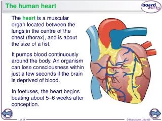
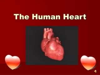
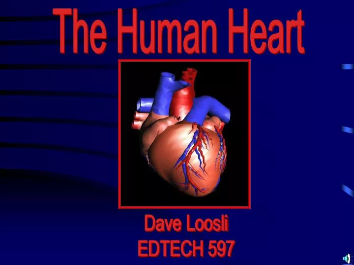
The Human Heart. Dave Loosli EDTECH 597. General Anatomy. Regions of the Heart Base and Apex (point of heart) Left Ventricle Right Heart Pulmonary Pump (blood to lungs) Left Heart Systematic Pump (pressures of 100-120 mmHg). Right Heart Doesn’t extend into the apex Thinner walls.

E N D
The Human Heart Dave Loosli EDTECH 597
General Anatomy • Regions of the Heart • Base and Apex (point of heart) • Left Ventricle • Right Heart • Pulmonary Pump (blood to lungs) • Left Heart • Systematic Pump (pressures of 100-120 mmHg)
Right Heart Doesn’t extend into the apex Thinner walls Left Heart Extends into the apex Thicker walls Higher pressure Difference between the Right and Left Hearts
Right Heart Doesn’t extend into the apex Thinner walls Left Heart Extends into the apex Thicker walls Higher pressure Difference between the Right and Left Hearts True or False: The Right Heart has thicker walls than the Left Heart? FALSE The Left Heart has thicker walls.
Tissue Layers of the Heart • Endocardium • Myocardium • Pericardium • visceral pericardium (epicardium) • parietal pericardium (pericardial sac) • pericardial fluid) between pericardial sac and epicardium)
Tissue Layers of the Heart • Endocardium • Myocardium • Pericardium Name the three layers of the heart
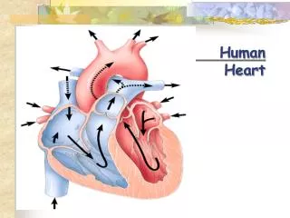
Pathway of Blood Flow through the Heart(Unoxygenated) • Inferior Vena Cava and Superior Vena Cava • (also coronary sinus = the venous return from coronary veins) • Right Atrium and Right Auricle • Tricuspid Valve • chordae tendonae • tendon cords attached to tricuspid valve to prevent blood from going back into the atrium • papillary muscle • anchors valve • Right Ventricle • Pulmonary Semilunar Valves • Pulmonary Artery • Pulmonary Circulation • (arteries, arterioles, capillaries, venules, veins) -- exchanges of O2 and CO2
Pathway of Blood through the Heart(Oxygenated) • Pulmonary Veins • carry O2 back to the heart from the lungs. • Left Atrium and Left Auricle • Bicuspid (mitral) Valve • chordae tendonae • tendon cords attached to mitral valve to prevent blood from from going back into the atrium • papillary muscle • anchors valves • Left Ventricle • Aortic Semilunar Valves • Aorta • Coronary Arteries
The “Lub-Dub” of the Heart • Aortic Semilunar Valve (closed) • Pulmonary Semilunar Valve (closed) • Tricuspid Valve • Bicuspid Valve • Semilunar Valves )open) • Tricuspid and Bicuspid (mitral) Valves (closed) • Semilunar Valve (closed • Tricuspid and Bicuspid (mitral) Valves (open)
EKG Explanation • P wave • result of action potentials that cause depolarization of the atrial myocardium, signals the onset of atrial contraction. • QRS wave • composed of 3 individual waves-- Q, R, S waves • QRS complex results from ventricular depolarization and signals the onset of ventricular contraction. • T wave • represents the repolarization of the ventricles and precedes ventricular relaxation.
EKG Explanation • P wave • QRS wave • T wave What are the three waves of the EKG?
Arrythmias What are the TWO types of ARRYTHMIA? Tachycardia and Bradycardia How do you describe BRADYCARDIA? How do you describe TACHYCARDIA?
Heart Blocks • First Degree block • prolonged AV conduction • (prolonged P-R [or P-Q] interval) • Second Degree block • mobitz type I and II • (2:1, 3:1 or 4:1 rhythm) • Third Degree block • complete AV block • no synchrony between atria and ventricles (no synchrony between the P and QRS waves) • “ventricular escape’ • some site will take over as ventricular site