Download
1 / 24
240 likes | 342 Vues
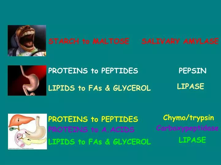
What happens where?. REACTANT/PRODUCT. ENZYME. STARCH to MALTOSE. SALIVARY AMYLASE. PROTEINS to PEPTIDES. PEPSIN. LIPASE. LIPIDS to FAs & GLYCEROL. Chymo/trypsin. PROTEINS to PEPTIDES. Carboxypeptidase. PROTEINS to A.ACIDS. LIPASE. LIPIDS to FAs & GLYCEROL. MALTASE.

E N D
What happens where? REACTANT/PRODUCT ENZYME STARCH to MALTOSE SALIVARY AMYLASE PROTEINS to PEPTIDES PEPSIN LIPASE LIPIDS to FAs & GLYCEROL Chymo/trypsin PROTEINS to PEPTIDES Carboxypeptidase PROTEINS to A.ACIDS LIPASE LIPIDS to FAs & GLYCEROL
MALTASE MALTOSE to GLUCOSE SUCRASE SUCROSE to GLUCOSE + FRUCTOSE LACTASE LACTOSE to GLUCOSE + GALACTOSE PEPTIDASE PEPTIDES to AMINO ACIDS
MUCOSA – layer closest to the lumen. First layer is epithelial cells with goblet cells that secrete a mucus to protect epithelium from enzymes. Beneath this is connective tissue 4 LAYERS ! SUBMUCOSA – made up of Connective tissue. Here Blood vessels and nerves lie & fibrous proteins (elastin) LUMEN MUSCULARIS EXTERNA – Two bands of muscle lie here (longitudinal & circular).- this aids peristalsis & mixes food. SEROSA – thin layer of Connective tissue
We are going to look at the following areas of the digestive system in more detail: The mouth and the oesophagus The stomach The liver and pancreas The small intestine The colon
C & L muscles Help to swallow Food. Bolus enters the Stomach. MASTICATION – chewing food using molars & premolars. 3 pairs of Salivary glands release saliva. Soluble materials dissolve. Starch to Maltose (S.Amylase)
Cardiac sphincter – opens To let bolus into stomach Pyloric sphincter – opens to let bolus into the duodenum CHYME
A Gastric Pit Creates a very folded surface which secretes gastric juices. Creates a very folded surface which secretes gastric juices. Gastric juices are approx. pH 1.
A Gastric Pit Parietal (oxyntic) cells release HCL. pH 1 kills lots of Bacteria. Chief cells release Pepsinogen. Pepsinogen is an inactive enxyme
A Gastric Pit HCL + pepsin work together to convert inactive PEPSINOGEN to PEPSIN Gastric juice also contains gastric LIPASE Gastric mucus is produced to protect epithelium from low pH Little absorption occurs in the stomach
LIVER BILE DUCT GALL BLADDER STOMACH PANCREAS THE DUODENUM – PANCREAS & LIVER
Pyloric sphincter relaxes – chyme leaves the stomach. The liver prdc. bile This is stored in the gall bladder. Bile moves down the bile duct and into the Duodenum. THE DUODENUM – PANCREAS & LIVER
What is bile? Salts: Sodium glycocholate Sodium taurocholate Salts are derived from cholesterol Ions: Hydrogencarbonate ions THE DUODENUM – PANCREAS & LIVER
Salts These emulsify fats Hydrogencarbonate ions These neutralise the acidic Chyme from the stomach pH 1 pH 7 Droplets to single fats
Pancreas has a dual function…… ……as an ENDOCRINE & EXOCRINE gland THE DUODENUM – PANCREAS & LIVER PANCREAS
Pancreas as an ENDOCRINE gland. Pancreatic juice is Made in the pancreas and secreted into the duodenum Enzymes: TRYPSIN CHYMOTRYPSIN ENTEROKINASE LIPASE AMYLASE CARBOXYPEPTIDASE
TRYPSIN & CHYMOTRYPSIN - are both proteases They are initially release in their inactive forms TRYPSINOGEN CHYMOTRYPSINOGEN ENTEROKINASE catalyses the following reactions TRYPSINOGEN to TRYPSIN CHYMOTRYPSINOGEN to CHYMOTRYPSINOGEN Pancreatic juice also contains HYDROGENCARBONATE ions, this keeps the pH at NEUTRAL.
THE SMALL INTESTINE – • Composed of 3 parts: • DUODENUM (25cm • JEJUNUM • (2m) • (3) ILEUM • (2.75m) THE SMALL INTESTINE – 5m long
LUMEN Villi Crypts of Lieberkuhn Goblet cells Paneth cells THE SMALL INTESTINE – 5m long
One villus has lots of MICROVILLI MICROVILLI: 1 µm long and 0.1 µm wide
GOBLET CELL secretes mucus Artery, vein & lymphatic Capillary are all important In removing digested food.
Crypt of Lieberkuhn Goblet cells are found here So are ……….. PANETH cells. Phagocytosis?
Two types of digestion in the Small Intestine Occurs in the LUMEN Pancreatic juices continue to work. Occurs on the surface of VILLI. Many epithelial cells have enzymes embedded into their plasma membranes. Some enzymes become adsorbed into the GLYCOCALYX
The final products of digestion are: AMINO ACIDS FATTY ACIDS GLYCEROL MONOSACCHARIDES We have moved from large insoluble polymers to small soluble monomers All of these products then cross the plasma membrane of the villi and then enter blood lymphatic capillaries Diffusion, active transport and facilitated diffusion all have a role