Download
1 / 23
290 likes | 1.16k Vues
Lecture Notes Series I. 2008 Cell Signaling Biology 4407B, Biochemistry 4806B, Neuroscience 4376B Pharmacology 5409B. G protein Coupled Receptors and G proteins. . . Dr. Melanie Kelly, Department of Pharmacology, Dalhousie University. mkelly@dal.ca.

E N D
Lecture Notes Series I. 2008 Cell SignalingBiology 4407B, Biochemistry 4806B, Neuroscience 4376B Pharmacology 5409B G protein Coupled Receptors and G proteins . Dr. Melanie Kelly, Department of Pharmacology, Dalhousie University. mkelly@dal.ca
G proteins Couple Receptors to Effectors • Receptors are Seven Transmembrane Serpentine receptors • Receptors are specialized for interaction with G proteins • Effectors are enzymes (Phospholipases, Kinases, Synthases) and other target molecules such as ion channels.
GPCR Superfamily • 1983 rhodopsin was the first G-protein-coupled receptor to be cloned and was found to have seven hydrophobic segments, constituting 7 transmembrane segments. • 7TM receptors now known to be the largest superfamily of proteins in eucaryotes. • GPCRs signal via coupling to guanine nucleotide binding proteins. • 7 TM receptors may exist as oligomeric protein structures and linked to multiple G-proteins to initiate pleiotropic effects.
G-Protein-Coupled Receptors Have Evolved Through Divergent and Convergent Evolution 20% 70% 70%
Rhodopsin- The Founding member of Family A! • Members of Family A constitute aprox. 90% of all GPCRs. • Rhodopsins are activated by light and turn on signaling pathways that leads to vision.
Structural Characteristics of Rhodopsin Family • Conserved residues largely located in transmembrane segments are a “finger-print signature” of the Rhdopsin-like GPCR family
7TM Receptor Family B • Family B is a 7 TM family of peptide and neurohormone receptors thatdo not have the identifying conserved residues of the rhodopsin-like receptors. • Family B have a large N-terminal extracellular domain containing 6 conserved cysteine residues. • All receptors in this family stimulate AC and couple to Gs proteins. • Some members of family B interact with chaperone proteins called RAMPs (receptor activity modifying proteins).
7TM Receptor Family C • Contains receptors for metabotropic glutamate receptors, Ca2+-sensing receptor (CaSR), the -aminobutyric acid (GABA)B receptor, some pheromone receptors and taste receptors. • Most family 3 GPCRs have a large extracellular domain responsible for ligand recognition and all Family C receptors function as dimers.
Guanine Nucleotide Binding Regulatory Proteins • GTP binding proteins act as transducers between receptors and effectors • Two major classes of G proteins have been described in the literature: Heterotrimeric G proteins consisting of , subunits and low molecular weight monomeric G proteins. • * subunits of trimeric G proteins (39-52 kDa) characterizes the individual G protein and have a high affinity Mg2+ -modulated nucleotide binding site. • * subunits are 35-36 kDa and the -subunit is 7-16 kDa. • * Low-molecular weight monomeric G proteins are 18-32 kDa and include ras-related proteins and many oncogene products.
Heterotrimeric G Protein Diversity • G proteins derived from large gene family at least 16 genes encode -subunit, 4 genes encode -subunits and multiple genes encode -subunit. • A given subtype may have more than one isotype: Gs includes 4 isotypes one of which is Golf Gi consists of Gi1,G i2, G i3 Gq consists of G15, G16, G14, G11, Gq G12 consists of G12 and G13 • In mammals 5 distinct -subunits and several different -subunits have been identified.
Interactions between G proteins and Receptors (a) Agonist binds to receptor (b) G protein interacts with activated receptor and GDP exchanged for GTP on subunit and subunits dissociate (?) (c) Activated subunit interacts with other signaling molecules. subunits also signal. (d) -subunit possess slowly hydrolysing intrinsic GTPase activity. RGS proteins enhance hydrolysis of GTP to GDP. (e) -subunit inactivated and recycled back to associatewith subunits.
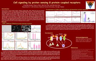
Agonists and Antagonists Act by Stabilizing Active and Inactive Receptor Conformations • 7TM Receptors are in equilibrium between active and inactive conformations. -Dynamic allosteric proteins that interchange between different conformations that are stabilized by ligands • An agonist -stimulates the receptor by stabilizing an active conformation by various mechanisms including binding between transmembrane segments • An antagonist is a compound that stabilizes one or more of the many different inactive conformations of the receptor and thus prevents the receptor from going into the active signaling conformation.
Desensitization Mechanisms Turn Signaling Off During Prolonged Stimulation • G protein-coupled receptor signalling is attenuated by phosphorylation by protein kinases and interaction with an intracellular protein, arrestin, which binds to the phosphorylated receptor and promotes dissociation of G protein • Ligand-occupied receptors are then sequestered into endocytic intracellular vesicles where the ligand is degraded and the receptor dephosphorylated and shunted back to the membrane. • After repeated or prolonged activation downregulation, receptors can be delivered to lysosomes and degraded after internalization. This results in a decreased response which is only recoverable with synthesis of new receptor molecules.
Kinases and Arrestins involved in desensitization and internalization of GPCRs • GRKs – G protein receptor kinases – homologous desensitization. • Protein Kinase C and Protein Kinase A – heterologous desensitization.
Some G protein-coupled Receptors Activate cAMP-dependent Signaling Pathways
Some G protein-coupled Receptors Activate Phospholipid Signaling Pathways
G Protein Receptor Cross-Talk for Signal Amplification Selbie and Hill, 1998 TIPS 19: 87-93
Bacterial Toxins Can Regulate The Activity of Specific G protein Subunits • Several bacterial toxins modify G protein activity by a process called ADP-ribosylation. This process involves the addition of an ADP-ribose group from nicotinamide adenine dinucleotide (NAD+) to an amino acid residue in the subunit. • Cholera Toxin – ADP-ribosylates and irreversibly activates Gs by inhibiting its GTPase activity. This leads to prolonged pathway activation. • Pertussis Toxin - ADP-ribosylates and inactivates Gi and Go by stabilizing their association with subunits. This leads to inhibition of GPCR pathways that couple to Gi/o G proteins.