Download
1 / 92
980 likes | 1.57k Vues
HEART DISEASES. HEART DISEASES (HD). FIVE MAJOR TYPES Ischemic HD Hypertensive HD Valvular /Endocardial HD Cardiomyopathy HD Congenital HD Other s : pericarditis, tumor, et al. ISCHEMIC HEART DISEASE. Myocardial ischemia.

E N D
HEART DISEASES (HD) FIVE MAJOR TYPES Ischemic HD Hypertensive HD Valvular/Endocardial HD Cardiomyopathy HD Congenital HD Others: pericarditis, tumor, et al.
Myocardial ischemia Myocardial ischemia is a condition in which oxygen deprivation to the heart muscle is accompanied by inadequate removal of metabolites because of reduced blood flow or perfusion.
Ischemic Heart Disease Ischemic heart disease (IHD) is the generic designation for a group of closely related syndromes resulting from ischemia– an imbalance between the supplyand demand of the heart for oxygenated blood. .
An imbalance occurs between myocardial oxygen supply and demand.
“Supply” ischemia Acute reduction of oxygen supply secondary to increased coronary vascular tone (ie, coronary vasospasm) or by marked reduction or cessation of coronary flow as a result of platelet aggregates or thrombi. Responsible for myocardial infarction (MI) Most episodes of unstable angina (UA) In many circumstances, ischemia results from both an increase in oxygen demand and a reduction in supply.
“Demand" ischemia In the presence of coronary obstruction, an increase of myocardial oxygen requirements caused by exercise, tachycardia, or emotion leads to a transitory imbalance. Responsible for most episodes of chronic stable angina
Ischemia may manifest as (1) anginal discomfort, (2) ST-segment deviation on ECG, (3) reduced uptake of thallium 201 or technetium 99 in myocardial perfusion images, (4) regional or global impairment of ventricular function.
Determinants of myocardial oxygen consumption Heart rate Contractility Systolic wall tension Maintenance of cell viability in basal state Depolarization Activation Maintenance of active state Direct metabolic effect of catecholamines Fatty acid uptake
Ischemia comprises not only insufficiency of oxygen (hypoxia, anoxia), but also reduced availability of nutrient substratesand inadequate removal of metabolites.
Because coronary artery narrowing or obstruction owing to atherosclerosis underlies myocardial ischemia in the vast majority of cases, IHD is often termed coronary artery disease(CAD) or coronary heart disease(CHD).
The acute coronary syndromes of unstable angina, acute myocardial infarction, and sudden death share a common pathophysiologic basis, with coronary atherosclerotic plaque rupture as the pathologic hallmark and associated intraluminal platelet-fibrin thrombus.
The dominant influence in the causation of the IHD syndromes : Fixed atherosclerotic narrowing of the coronary arteries, Intraluminal thrombosis overlying a ruptured or fissured atherosclerotic plaque, Platelet aggregation, Vasospasm.
Role of Fixed Coronary Obstructions More than 90% of patients with IHD have advanced stenosing coronary atherosclerosis (“fixed” obstructions). Although only a single major coronary epicardial trunk may be affected, more often two or all threeare involved .
Role of Acute Plaque Change Acute myocardial ischemia is often precipitated by disruption of previously only partially stenosing atherosclerotic plaques with hemorrhage, fissuring, or ulceration. This is often followed by mural or total thrombosis. Such vascular injury is fundamental to the development of the acute coronary syndromes–unstable angina, acute myocardial infarction, or sudden ischemic death–in most patients. High-grade but slowly developing occlusions probably stimulate well-developed collateral vessels over time that may protect against infarction.
Role of Coronary Thrombus Coronary thrombosis, partial or total, plays a critical role in acute coronary syndromes. In myocardial infarction, the added thrombus converts a disrupted, partially stenotic plaque to a complete stenosis. Thrombus is a potent activator of multiple growth-related signals in smooth muscle cells; both platelet-mediated and smooth muscle cell-mediated events contribute to the growth of atherosclerotic lesions.
Role of Vasoconstriction Transient vasoconstriction may be induced at a site of plaque disruption and thrombosis.
Terminology: Ischemic Heart Disease • Ischemia - Insufficient blood supply injury Angina (reversible) or MI (irreversible) • Angina(Angina=Strangulation Pectoris=Chest) • Myocardial infarction (MI)- Death of myocardial cells (irreversible) • Cardiac failure: Impaired pumping ability of the heart • Cardiogenic shock: Shock due tocardiac failure • Arrhythmias:cardiac rhythm disturbances
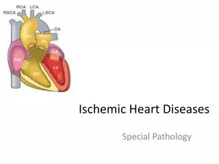
Coronary circulation Rich Blood supply High Oxygen consumption (A-V19-9ml/100ml) Two coronary arteries-225 ml/min Direct branch from aorta End arteries – few collaterals Phasic blood flow- during diastole Local metabolism is primary controller of coronary blood flow
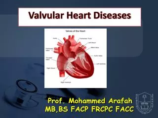
Coronary Arteries • Left Coronary Artery • Anterior Descending • Circumflex LCx LAD Right Coronary Artery
Abnormalities of O2 supply Increased O2 demand High heart rate, BP, Contractility Insufficient O2 supply Anemia and Hypoxia Coronary Vessels Intermittent (Spasm, Congenital) Permanent (Atherosclerotic Plaque)
Depending on the rate of development and ultimate severity of the arterial narrowing(s) and the myocardial response, four ischemic syndromes may result: (1) angina pectoris (2) myocardial infarction (3) chronic ischemic heart disease (4) sudden cardiac death Ischemic syndromes
1. AnginaPectoris Stable (classic, typical, Heberden) angina Unstable (pre-infarction, crescendo, acute coronary insufficiency) angina Prinzmetal's ("variant") angina
Anginas are caused by varying combinations of increased myocardial demand and decreased myocardial perfusion, owing to fixed stenosing plaques disrupted plaques vasospasm thrombosis platelet aggregation embolization
Angina pectoris is a symptom complex of IHD characterized by paroxysmal attacks of substernal or precordial chest discomfort (variously described as constricting, squeezing, choking, or knife-like) Caused by transient (15 seconds to 15 minutes) myocardial ischemia that falls short of inducing the cellular necrosis that defines infarction.
Stable (classic, typical, Heberden) angina The pathogenesis of typical angina pectoris appears to be the reduction of coronary perfusion to a critical level by chronic stenosing coronary atherosclerosis. This renders the heart vulnerable to further ischemia whenever there is increased demand, such as that produced by physical activity, emotional excitement, or any other cause of increased cardiac workload. Relieved by restor nitroglycerin, a strong coronary vasodilator (thereby increasing supply).
Unstable (pre-infarction, crescendo, acute coronary insufficiency) angina Refers to a pattern of pain that occurs with progressively increasing frequency, is precipitated with progressively less effort, often occurs at rest, and tends to be of prolonged duration. In most cases, this is probably due to a thrombus developing over a ruptured plaque.
In unstable angina, a relatively small fissure or disruption of an atherosclerotic plaque may lead to a sudden change in plaque morphology, with platelet aggregation or mural thrombus and frequently vasoconstriction leading to transient reduction in coronary blood flow. Untreated, many of these people get an MI soon.
Prinzmetal's ("variant") angina This is primarily attributable to vasospasm. Perhaps it's the cardiac equivalent of migraine. Although individuals with this form of angina may well have significant coronary atherosclerosis, the anginal attacks are generally unrelatedto physical activity, heart rate, or blood pressure. Prinzmetal’s angina generally responds promptly to vasodilators, such as nitroglycerin and calcium channel blockers.
2. Myocardial Infarction “Heart attack” • Death of Myocardial muscle due to lack of blood supply. • Most common cause is Atherosclerotic narrowing of coronary arteries.
Pain in Myocardial Infarction Precordial , intense, constrictive Similar to Angina – severe prolonged Radiates to shoulder and left arm May present in other location-jaw, epigastrium. Often with breathlessness, nausea, vomiting & perspiration Hypotension Collapse Less severe or absent in elderly No response to nitrates
Causes of myocardial infarcts Atherosclerosis:Makes up 90% of coronary artery disease Cocaine:The second most common cause of myocardial infarction and sudden cardiac death Prinzmetal's coronary spasm Vasculitis: (1) lupus; (2) polyarteritis nodosa; (3) rheumatoid arthritis; (4) Kawasaki's; (5) Takayasu's; (6) mycotic aneurysms. Embolization Syphilis(mesaortitis syphilitica) Dissecting hematoma Shock and left-sided failure (subendocardial infarct)
There are two types of myocardial infarction, each having differing morphology and clinical significance: (1) The transmural infarct (more common type) (2) The subendocardial (nontransmural) infarct
The transmural infarct The ischemic necrosis involves the full or nearly full thickness of the ventricular wall in the distribution of a single coronary artery. This pattern of infarction is usually associated with coronary atherosclerosis plaque rupture superimposed thrombosis
Subendocardial Infarction An area of ischemic necrosis limited to the inner one-third or at most one-half of the ventricular wall, often extending laterally beyond the perfusion territory of a single coronary artery. There is diffuse stenosing coronary atherosclerosis and global reduction of coronary flow but neither plaque rupture nor superimposed thrombosis.
Coronary Arterial Occlusion At least 90% of transmural acute myocardial infarcts are caused by an occlusive intracoronary thrombus overlying an ulcerated or fissured stenotic plaque. Every acute transmural myocardial infarct, a dynamic interaction has occurred among several or all of the following: severe coronary atherosclerosis, an acute atheromatous plaque change (fissuring, ulceration), superimposed thrombosis, platelet activation, and vasospasm.
In the typical case, the following sequence of events can be proposed (90%): Sudden change in the morphology of an atheromatous plaque, i.e., intraplaque hemorrhage, ulceration, or fissuring. Plateletadhesion, aggregation, activation, and release of adenosine diphosphate (ADP), with buildup of a platelet mass. The platelet mass may give rise to emboli or potentiate occlusive thrombosis. Tissue thromboplastin is released. Adherent activated platelets release thromboxane A2, serotonin, and platelet factors 3 and 4 (predisposing to coagulation, favoring vasospasm). Frequently within minutes, the thrombus evolves to become completely occlusive.
In the approximately 10% of cases In some of the cases, the coronary arteries are free of atherosclerosis by angiography: Vasospasm with or without coronary atherosclerosis may induce the acute perfusion deficit, perhaps in association with platelet aggregation. Emboli from a left-sided mural thrombosis or vegetative endocarditis or paradoxic emboli from the right side of the heart or the peripheral veins (through a patent foramen ovale) could cause coronary occlusion.
Coronary Atheorsclerosis LCx LAD Right Coronary Artery
Myocardial Infarction : Myocardial Response Occlusion of a major coronary artery results in ischemia throughout the anatomic region supplied by that artery (called area at risk), Acutely ischemic myocardium undergoes progressive biochemical, functional, and morphologic changes, the outcome of which largely depends on the severity and duration of flow deprivation.
The principal biochemical consequence of myocardial ischemia is the onset of anaerobic glycolysis within seconds, leading to inadequate production of high-energy phosphates (e.g., creatine phosphate and adenosine triphosphate [ATP ]) and accumulation of potentially noxious breakdown products (such as lactic acid). Myocardial function is exceedingly sensitive to severe ischemia; striking loss of contractility is evident within 60 seconds of onset.
Reversible ultrastructural changes develop within few minutes after onset of ischemia e.g., cell and mitochondrial swelling, glycogen depletion. Although function becomes strikingly abnormal within few minutes after onset of ischemia, myocardial coagulation necrosis occurs only after 20 to 40 minutes of severe ischemia.