Download
1 / 1
10 likes | 79 Vues
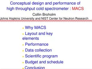
Progress on the New High Intensity Cold Neutron Spectrometer, MACS C. Broholm 1,2 , T. D. Pike 1,2 , P. K. Hundertmark 1,2 , P. C. Brand 2 , J. W. Lynn 2 , D. J. Pierce 2 , J. Moyer 2 , J. G. LaRock 2 , S. A. Smee 1 , G. Scharfstein 1 , R. Hammond 1 , and J. Orndorff 1. Shutter. Collimators.

E N D
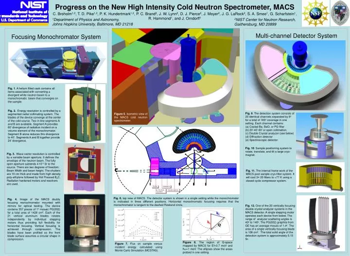
Progress on the New High Intensity Cold Neutron Spectrometer, MACS C. Broholm1,2, T. D. Pike1,2, P. K. Hundertmark1,2, P. C. Brand2, J. W. Lynn2, D. J. Pierce2, J. Moyer2, J. G. LaRock2, S. A. Smee1, G. Scharfstein1, R. Hammond1, and J. Orndorff1 Shutter Collimators Variable aperture Figure 7. Flux on sample versus incident energy calculated using Monte Carlo Simulation (MCSTAS). kf Be, PG, Al2O3 Cryo filters ki Q Figure 8. The region of Q-space mapped by MACS for Ef=3.7 meV and ħw=1 meV. The ellipses show the areas probed in one setting. 1Department of Physics and Astronomy, Johns Hopkins University, Baltimore, MD 21218 2NIST Center for Neutron Research, Gaithersburg, MD 20899 Multi-channel Detector System Focusing Monochromator System Fig. 1. A helium filled cask contains all items associated with converting a divergent white neutron beam to a monochromatic beam that converges on the sample. Fig. 2. Energy resolution is controlled by a segmented radial collimating system. The blades of the device converge at the center of the cold source. Two in-line segments A and B are available. Segment A provides 60’ divergence of radiation incident on a volume element of the monochromator. Segment B alone reduces this divergence to 40’. Segments A and B together provide 24’ divergence. Fig. 9. The detection system consists of 20 identical channels separated by 8o for a total of 160o coverage in one setting. Each channel contains: (a) Cooled Be, BeO, or PG filter (b) 20’-40’-60’ or open collimation. (c) Double Crystal analyzer (see below) (d) Diffraction detector (e) Spectroscopic detector Figure 5. Isometric view of the MACS cold neutron spectrometer. Fig. 10. Sample positioning system to rotate, translate, and tilt a large cryo-magnet. Fig. 3. Wave vector resolution is controlled by a variable beam aperture. It defines the envelope of the neutron beam. The fully open aperture subtends 4.10-3 Sr to the source. There are two degrees of freedom: Beam Width and beam height. The shutters are 10 cm thick and made from high density poly-ethylene followed by Hot Pressed B4C. Radiation hardened motors and resolvers are used. Fig. 11. The internal frame work of the MACS post sample cryo-filter system. It will cool 3× 20 filters to <77 K using a closed cycle compressor system. Fig. 6. top view of MACS. The detector system is shown in a single setting while the monochromator is indicated in three different positions. Horizontal monochromatic focusing requires that the monochromator is tangent to the dashed Rowland circle. Fig. 4. Image of the MACS doubly focusing monochromator mounted with mirrors for optical testing. The device contains 357 pieces of 1o mosaic PG(002) for a total area of 1428 cm2. Each of the 21 vertical aluminum blades rotates independently by individual stepping motors thus providing full flexibility for horizontal focusing. Vertical focusing is achieved through compression. The blades have been profiled so the front blade surface assumes a circular shape in compression. Fig. 12. One of the 20 vertically focusing double crystal analyzer systems in the MACS detector. A single stepping motor operates each device from below. The range of analyzer scattering angles is 40o to 140o. The PG(002) graphite from GE has an average mosaic of 1.4o. The area of a single vertically focusing blade is 108 cm2. The total solid angle of the detection system is approximately 0.15 Sr.