Download
1 / 10
100 likes | 257 Vues
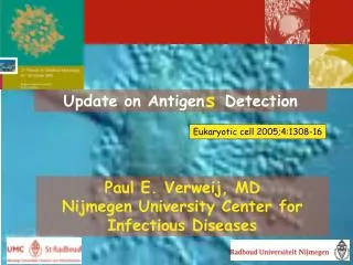
Detection of Carcinoembryonic antigen with sandwich-biosensor. Alexander Pak, Antonina Malozemova , Luiza Niyazmetova, Mukhtar Sadykov , Zeinaf Muradova Nazarbayev University, School of Science and Technology, Department of Biological Sciences. Introduction.

E N D
Detection of Carcinoembryonic antigen with sandwich-biosensor Alexander Pak, AntoninaMalozemova, Luiza Niyazmetova, MukhtarSadykov, ZeinafMuradovaNazarbayev University, School of Science and Technology, Department of Biological Sciences
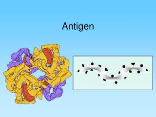
Introduction Figure 1. Carcinoembryonic antigen Carcinoembryonic antigen (CEA) is one of the examples of the biomarker which appears at early stages of such types of cancer as colorectal, gastric, pancreatic, lung, and breast carcinoma [1]. Aims: • To obtain aptamers against carcinoembryonic antigen (CEA) • To express streptavidin on yeast and bacterial cell surfaces • To make sandwich-biosensor to detect different types of cancer at early stages
Aptamers and SELEX Figure 2. SELEX procedure Figure 3. SELEX 11 Figure 4. SELEX 12 gel run Aptamers – short synthetic oligonucleotides that have high specificity of interaction with a particular target[2] SELEX – Systematic Evolution of Ligands by Exponential enrichment. 12 cycles of SELEXwere finished Characterization step of aptamers from cycles 11 and 12 is ongoing Characterization is done on SPR (Surface Plasmon Resonance), which determines kinetics and affinity of aptamers for CEA.
Future plans Sequence and Synthesis of aptamers Characterize them via SPR Construct the parts for E.Coli and S.Cerevisia
References 1) Cancer diagnosis – information about cancer. (2013). Stranford Cancer Center . Retrieved from cancer.stanford.edu/information/cancerDiagnosis/ 2) Mascini, M. (2009). Aptamers in bioanalysis. John Wiley and Sons 3) Lim, H. K., Hwang, I., Sh., Park. (2011). Biotin-Assisted Folding of Streptavidin on the Yeast Surface. American Institute of Chemical Engineers. Retrieved from: http://www.cbe.buffalo.edu/people/pdfPub.jsp?id=2887 4) Huang, D. & E., Shusta. (2005). Secretion and Surface Display of Green Fluorescent Protein Institute of Chemical Engineers. Vol 21 No2. (349-357): Retrieved from: http://46.38.63.192/mail/lg.php?doi=10.1021/bp0497482&url=aHR0cDovL2xpYmdlbi5vcmcvc2NpbWFnMy8xMC4xMDIxL2JwMDQ5NzQ4Mi5wZGY%3D 5) Tan, A., Yildirimer, L., Rajadas, J., De La Peña, H., Pastorin, G., Seifalian, A. (2011). Quantum Dots and Carbon Nanotubes in Oncology.Nanomedicine. 6(6):1101-1114. Retrieved fromhttp://www.medscape.com/viewarticle/749698_2