Download
1 / 54
550 likes | 615 Vues
Cell Structure and Function. Chapter 7. Life is Cellular. The Cell Theory 1500’s – Europeans used lens to look at cloth quality 1600’s – two useful instruments were made with lens from the 1500’s Microscope Telescope Leeuwenhoek was the first to study nature under a microscope

E N D
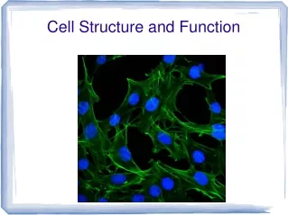
Cell Structure and Function Chapter 7
Life is Cellular • The Cell Theory • 1500’s – Europeans used lens to look at cloth quality • 1600’s – two useful instruments were made with lens from the 1500’s • Microscope • Telescope • Leeuwenhoek was the first to study nature under a microscope • 1665 – Hooke used the first light microscope to look at plant tissue • Looked at cork • Found the cells are the basic unit of life
Life is Cellular • Cell Theory • 1838 – Schelldon found all plants are made of cells • 1829 – Schwann found that all animals are made of cells • 1855 Virchow found cells come from cells • 1931 – Plowe found cells have cell membrane that is a physical structure between cells • 1970 – Maegulis proposes theory of tiny organelles in the cell once were free living cells
Life is Cellular • Cell theory states • All living things are composed of cells • Cells are the basic structure and function in living things • New cells are produced from existing cells
Life is Cellular • Cells come in many shape and sizes • Typical cell 5 to 50 micrometers in diameter • The tiniest bacteria are only 0.2 micrometer across and hard to see with the most powerful light microscope • Chaos chaos a giant bacteria is 1000 micrometers and can be seen with naked eye • Common structures in most cells • Cell membrane • Cell wall • Nucleus • cytoplasm
2 categories of cells Prokaryotes – smaller and simpler cells No nuclear membrane (genetic material dispersed throughout cytoplasm) Cell membrane prokaryotic DNA is circular (it has no ends). Cytoplasm No membrane-bound organelles All bacteria Escherichia coli lives in your intestine Carries out every activity associated with life Grow Reproduce Respond to stimulus Move sometime during their life cycle
Eukaryotes Contain a nucleus, cell membrane, and cytoplasm Most contain organelles Specialized structures that perform important cellular functions Some single cell organism Most are Multicellular organisms Eukaryotic DNA is linear
Eukaryotic Cell Structure Organelles – little organs Structures that are specialized for certain processes 2 major parts of a cell Nucleus Cytoplasm
Eukaryotic Cell Structure Cytoplasm – Cytoplasm is basically the substance that fills the cell. It is a jelly-like material that is eighty percent water and usually clear in color. It is more like a viscous (thick) gel than a watery substance, but it liquefies when shaken or stirred. Cytoplasm, which can also be referred to as cytosol, means cell substance. The cytoplasm is the site where most cellular activities occur, such as many metabolic pathways like glycolysis, and processes such as cell division.
Eukaryotic Cell Structure Nucleus - also sometimes referred to as the "control center", is a membrane-enclosed organelle found in eukaryotic cells. It contains most of the cell's genetic material, organized as multiple long linear DNA molecules in complex with a large variety of proteins, such as histones, to form chromosomes. Nuclear envelope - a double membrane that encloses the entire organelle and separates its contents from the cellular cytoplasm and is dotted with pores which allow material to flow in and out Chromatin – granular material consisting of DNA bound to a protein.
Eukaryotic Cell Structure Nucleus Chromosomes – condensed chromatins Happens when cells divide Distinct threadlike structures containing genetic information Nucleolus – small, dense region where ribosomes are formed
Eukaryotic Cell Structure Ribosomes - the components of cells that make proteins from amino acids. Small particles of proteins and RNA found throughout the cytoplasm Considered a small machine in a large factory producing proteins Cells active in producing proteins are often packed with ribsomes
Eukaryotic Cell Structure Endoplasmic Reticulum – responsible for the production of the protein and lipid components of most of the cell's organelles. The ER contains a great amount of folds - but the membrane forms a single sheet enclosing a single closed sac. This internal space is called the ER lumen. The ER is additionally responsible for moving proteins and other carbohydrates to the Golgi apparatus, to the plasma membrane, to the lysosomes, or wherever else needed.
Eukaryotic Cell Structure Endoplasmic Reticulum – There are two types of ER - rough, which is coated with ribosomes, and smooth, which isn't. Rough ER is the site of protein synthesis. The smooth ER is where the vesicles carrying newly synthesized proteins (from the rough ER) are budded off Uses enzymes to in the synthesis of lipid membranes and detoxification of drugs The liver cells play a role in detoxification and contain large amounts of ER
Eukaryotic Cell Structure Golgi Apparatus – The Golgi complex contains a great number of vesicles. These vesicles are used to send molecules to the cellular membrane, where they are excreted. There are also larger secretory vesicles, which are used for selective excretion. The Golgi is principally responsible for directing molecular traffic in the cell nearly all molecules pass through the Golgi complex at some point in their existence. The sorting is mediated by the vesicles. When proteins bind with their appropriate receptor on the vesicle, they are encoated in the vesicle and transported away.
Eukaryotic Cell Structure Lysosome The lysosome is a membranous bag which contains hydrolytic enzymes that are used to digest macromolecules. The lysosome contains over 40 enzymes
Eukaryotic Cell Structure Vacuoles – used only in plant cells. It is responsible for maintaining the shape and structure of the cell. Plant cells don't increase in size by expanding the cytoplasm, rather they increase the size of their vacuoles. The vacuole is a large vesicle which is also used to store nutrients, metabolites, and waste products. The pressure applied by the vacuole, called turgor, is necessary to maintain the size of the cell.
Eukaryotic Cell Structure Chloroplast – The chloroplast, basically, is the organelle responsible for photosynthesis. Structurally it is very similar to the mitochondrion. It contains a permeable outer membrane, a less permeable inner membrane, a intermembrane space, and an inner section called the stroma. However, the chloroplast is larger than the mitochondria Unlike other organelles, chloroplast and mitochondria have their own DNA.
Eukaryotic Cell Structure Mitochondria – Converts chemical energy stored in food into compounds that are more convenient for the cell to use The powerhouse of the cell The mitochondrion consists of four major sections – the outer membrane, the intermembrane space, the inner membrane, and the matrix.
Eukaryotic Cell Structure Mitochondria – The Outer Membrane -This membrane contains a great number of large transport proteins, which allows for large molecules to enter with ease. This membrane includes proteins that can convert lipid substrates into forms that can be used by the matrix. Inner membrane space - This space contains enzymes Inner membrane - This membrane is highly convoluted, forming many folds called cristae. This serves to greatly increase the surface area, allowing more work to be done is a smaller space Matrix - The Krebs Cycle takes place here. It also contains several copies of the mitochondrial DNA genome, special mitochondrial ribosome's, tRNAs, and various enzymes required for the expression of the mitochondrial gene
Eukaryotic Cell Structure Cytoskeleton – Eukaryotic cells have a wide variety of distinct shapes and internal organizations. Cells are capable of changing their shape, moving organelles, and in many cases, move from place to place. This requires a network a protein filaments placed in the cytoplasm known as the cytoskeleton. The two most important protein filaments are called the actin filaments and the microtubules. The actin is responsible for contraction (like in muscles) and the microtubules are for structural strength
Cell Boundaries Cell membrane – cell membrane is a flexible lipid bilayer. The lipid molecules (mostly phospholipids) that make up the membrane have a polar, hydrophilic head and two hydrophobic hydrocarbon tails. When the lipids are immersed in an aqueous solution the lipids spontaneously bury the tails together and leave the hydrophilic heads exposed. Thus this is a handy membrane to use, because it can automatically fix itself when torn
Cell Boundaries Cell Walls – It provides the most significant difference between plant cells and other eukaryotic cells. The cell wall is rigid (up to many micrometers in thickness) and gives plant cells a very defined shape. The cell wall is composed of cellulose fiber, polysaccharides, and proteins. In new cells the cell wall is thin and not very rigid. This allows the young cell to grow. This first cell wall of these growing cells is called the primary cell wall. When the cell is fully grown, it may retain its primary wall, sometimes thickening it, or it may deposit new layers of a different material, called the secondary cell wall.
Cell Boundaries Diffusion - the process by which molecules spread from areas of high concentration, to areas of low concentration. When the molecules are even throughout a space - it is called EQUILIBRIUM Concentration gradient - a difference between concentrations in a space. Equilibrium – concentration throughout a solution is the same Diffusion depends upon random particle movements, substances diffuse across membranes without requiring the cell to use energy
Cell Boundaries Osmosis – The diffusion of water (across a membrane) through a selectively permeable membrane Water will move in the direction where there is a high concentration of solute and hence a lower concentration of water.
Cell Boundaries Isotonic – A solution that contains the same concentration of solute as an another solution (e.g. the cell's cytoplasm). When a cell is placed in an isotonic solution, the water diffuses into and out of the cell at the same rate. The fluid that surrounds the body cells is isotonic.
Cell Boundaries Hypertonic solution – contain a high concentration of solute relative to another solution (e.g. the cell's cytoplasm). When a cell is placed in a hypertonic solution, the water diffuses out of the cell, causing the cell to shrivel
Cell Boundaries Hypotonic solution – contain a low concentration of solute relative to another solution (e.g. the cell's cytoplasm). When a cell is placed in a hypotonic solution, the water diffuses into the cell, causing the cell to swell and possibly explode.
Cell Boundaries Osmotic pressure – The pressure that must be applied to a solution to prevent the inward flow of water across a semi permeable membrane
Cell Boundaries Facilitated Diffusion – Glucose, sodium ions and chloride ions are just a few examples of molecules and ions that must get across the plasma membrane but to which the lipid bilayer of the membrane is virtually impermeable. Their transport must therefore be "facilitated" by proteins that span the membrane and provide an alternative route or bypass. Facilitated diffusion
Cell Boundaries Active Transport – Active Transport - When cells must move materials in an opposite direction - against a concentration gradient. It requires Energy. Proteins or Pumps are found in the cell membrane transport molecules across the membrane.
Cell Boundaries Molecular transport – Proteins are used to move small molecules such as calcium, potassium, and sodium ions across the membrane Endocytosis - cell takes in large particles by engulfing them Phagocytosis - "cell eating" - extensions off cytoplasm surround a particle and package it within a food vacuole and then the cell engulfs it. Ex. Amoebas use this process. Pinocytosis - the process of taking up liquid from the surrounding environment. Tiny pockets form along the membrane, fill with liquid, and pinch off. Exocytosis - cell gets rid of particles, opposite of endocytosis