Download
1 / 72
730 likes | 911 Vues
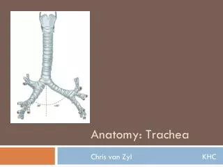
Trachea and esophagus. Ehab ZAYYAN, MD, PhD. Trachea. Cartilaginous and membranous tube Starts from the lower border of the cricoid cartilage (6 th cervical vertebra). In adults the trachea is about 11.25 cm long and 2.5 cm in diameter

E N D
Trachea and esophagus Ehab ZAYYAN, MD, PhD
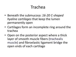
Trachea • Cartilaginous and membranous tube • Starts from the lower border of the cricoid cartilage (6th cervical vertebra)
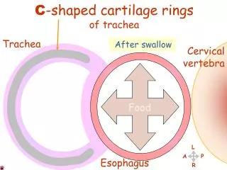
In adults the trachea is about 11.25 cm long and 2.5 cm in diameter • Ends at the (Carina) by dividing into two main bronchi at the level of the sternal angle (opposite to 4th and 5th thoracic vertebrae) • Consists of 16- 20 incomplete cartilage rings (U- shaped)
The fibroelastic tube is kept patent by the presence of U-shaped cartilaginous bar (rings) of hyaline cartilage embedded in its wall. The posterior free ends of the cartilage are connected by smooth muscle, thetrachealis muscle. • The mucous membrane of the trachea is lined with pseudostratified ciliated columnar epithelium and contains many goblet cells and tubular mucous glands.
Anterior relations of the trachea • Skin • Fascia • Sternothyroid and sternohyoid muscles • Isthmus of the thyroid • Inferior thyroid veins • Jugular arch • Thyroid ima artery • Left bracheoceohalic vein
Posterior relations • Right and left recurrent laryngeal nerves • Esophagus • Vertebral column
Lateral relations • Thyroid gland lateral lobes • Carotid sheath
Trachea in the neck Blood supply • The upper two thirds is supplied by the inferior thyroid arteries and the lower third is supplied by the bronchial arteries.
Lymphatic drainage • pretracheal and paratracheal lymph nodes
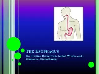
Esophagus • Muscular tube • 25 cm long • From the pharynx to stomach • Begins at the level of the cricoid cartilage, 6th cervical vertebra • It descends in the midline slightly to the left.
Relations of the esophagus in the neck • Anterior: trachea and RLN • Posterior: prevertebral fascia and vertebral column • Lateral: lobes of the thyroid and carotid sheath
Esophagus in the neck • Blood supply: inferior thyroid arteries and veins • Lymph: deep cervical lymph nodes • Nerves: RLN and sympathetic trunks
Midline structures in the neck • Symphysis menti • Submental triangle • Body of the hyoid bone: at the level of 3rd cervical vertebra • Thyrohyoid membrane • Upper border of thyroid cartilage: level of 4th cervical vertebra • Cricothyroid ligament • Cricoid cartilage: level of 6th cervical vertebra • Cricotracheal ligament • First ring of trachea • Isthmus of the thyroid gland • Thyroid ima artery • Jugular arch • Suprasternal notch
In young children, the thymus gland may extend above the suprasternal notch as far as the isthmus of the thyroid gland, and the brachiocephalic artery and the left brachiocephalic vein may protrude above the suprasternal notch.
Symphysis menti • Submental triangle • Hyoid bone body • Thyrohyoid membrane • Thyroid cartilage • Cricothyroid ligament • Cricoid cartilage • Cricotracheal ligament • First ring of trachea • Thyroid gland isthmus • Thyroid ima artery • Jugular arch • Suprasternal notch
Compromised airways Top Emergency !!!!!
Resuscitation A: Airways B: Breathing C: Circulation D: Drugs
Urgent airways management • Endotracheal intubation • Cricothyroidotomy • Tracheostomy
Cricothyroidotomy • Performed in top urgent situations • When endotracheal intubation is impossible • When there is no time even for tracheostomy • Incision is made through the skin, fasciae and cricothyroid membrane and a tube is inserted
Complications of cricothyroidotomy • Esophageal injury (young children!!) • Larynx injury • Hemorrhage
The history of tracheotomy refers to 200 years BC In 1800 the most common indication was laryngeal diphtheria Infectious causes of tracheotomy are decreasing while congenital causes are increasing Tracheostomy
Upper respiratory airway obstruction Pulmonary care Long ventilation Indications of tracheotomy
Congenital Craniofacial anomalies Laryngeal anomalies Bilateral vocal cord paralysis Tracheal anomalies Lymphatic anomalies Upper respiratory airway obstruction