Download
1 / 77
850 likes | 1.37k Vues
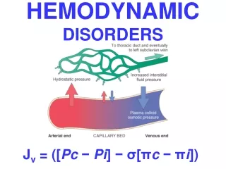
HEMODYNAMIC DISORDERS. Edema. 60% of lean body weight is water; two thirds of this water is intracellular, remainder is in the extracellular space, mostly interstitial fluid.

E N D
Edema 60% of lean body weight is water; two thirds of this water is intracellular, remainder is in the extracellular space, mostly interstitial fluid. EDEMA signifies increased fluid in the interstitial tissue spaces. Depending on the site, fluid collections are variously designated hydrothorax, hydropericardium, and hydroperitoneum (ascites). Anasarca: severe, generalized edema with profound subcutaneous tissue swelling.
Downloaded from: Robbins & Cotran Pathologic Basis of Disease (on 1 April 2005 06:43 PM) © 2005 Elsevier
Downloaded from: Robbins & Cotran Pathologic Basis of Disease (on 1 April 2005 06:47 PM) © 2005 Elsevier
Types of edema • Transdate: protein poor (<3 gm/dl) fluid with specific gravity of <1.012 due to imbalances in normal hemodynamic forces e.g. congestive heart failure, liver and renal disease etc. • Exudate - protein rich (>3 gm/dl) fluid with a specific gravity of >1.020 results from endothelial damage and alteration of vasular permeability e.g. inflammatory and immunologic pathology.
Hyperemia & Congestion • Hyperemia is an active process resulting from tissue inflow because of arteriolar dilation, e.g. skeletal muscle during exercise or at sites of inflammation. The affected tissue is redder because of the engorgement of vessels with oxygenated blood. • Congestion is a passive process resulting from impaired outflow from a tissue. It may be systemic e.g. cardiac failure, or local e.g. an isolated venous obstruction. The tissue has a blue-red color (cyanosis), due to accumulation of deoxygenated hemoglobin in the affected tissues.
Morphology: lung • The cut surfaces are hemorrhagic and wet. LUNGS: Microscopically, acute pulmonary congestion is characterized by alveolar capillaries engorged with blood,alveolar septal edema and/or focal intra-alveolar hemorrhage. In chronic pulmonary congestion, the septa are thickened and fibrotic, and the alveolar spaces may contain numerous hemosiderin-laden macrophages (heart failure cells).
Morphology: liver • In acute hepatic congestion: central vein and sinusoids are distended with blood with or without central hepatocyte degeneration. • In chronic passive congestion of the liver: on cut surface central regions of the hepatic lobules are red-brown and surrounded by zones of uncongested tan liver (nutmeg liver). Microscopically: centrilobular necrosis with loss of hepatocytes, hemorrhage and hemosiderin-laden macrophages. Long-standing cases (most commonly associated with heart failure), hepatic fibrosis (cardiac cirrhosis) may develope.
Hemorrhage • Hemorrhage generally indicates extravasation of blood due to vessel rupture • Hematoma: accumulation of blood within tissue. • Petechiae: Minute 1- to 2-mm hemorrhages into skin, mucous membranes, or serosal surfaces.
Purpura: Slightly larger (≥3 mm) hemorrhages. • Ecchymoses: Larger (>1 to 2 cm) subcutaneous hematomas (i.e., bruises). • Large accumulations of blood in one or another of the body cavities are called hemothorax, hemopericardium, hemoperitoneum, or hemarthrosis (in joints).
Thrombosis • It represents hemostasis in the intactvascularsystem. • It is a process by which a thrombus is formed. • A thrombus is a solid mass of blood constituents which developes in artery or vein. • Is intravascular coagulation of blood often causing sinificant interuption to blood flow.
Pathogenesis Three primary influences predispose to thrombus formation, the so-called Virchow triad: • endothelial injury • stasis or turbulence of blood flow • blood hypercoagulability In other words it results from interaction platelets, damaged endothelial cells and the coagulation cascade.
Figure 4-13 Virchow triad in thrombosis. Endothelial integrity is the single most important factor. Note that injury to endothelial cells can affect local blood flow and/or coagulability; abnormal blood flow (stasis or turbulence) can, in turn, cause endothelial injury. The elements of the triad may act independently or may combine to cause thrombus formation. Downloaded from: Robbins & Cotran Pathologic Basis of Disease (on 1 April 2005 07:37 PM) © 2005 Elsevier
Primary (Genetic) Hypercoagulable States • Mutation in factor V gene (factor V Leiden) • Mutation in prothrombin gene • Mutation in methyltetrahydrofolate gene • Antithrombin III deficiency • Protein C deficiency • Protein S deficiency • Fibrinolysis defects )
Secondary(Acquired)Hypercoaguable States High risk for thrombosis • Prolonged bed rest or immobilization • Myocardial infarction,Atrial fibrillation • Tissue damage (surgery, fracture, burns) • Cancer • Prosthetic cardiac valves • Disseminated intravascular coagulation • Heparin-induced thrombocytopenia • Antiphospholipid antibody syndrome (lupus anticoagulant syndrome) Lower risk for thrombosis Cardiomyopathy,Nephrotic syndrome,Hyperestrogenic states (pregnancy),Oral contraceptive use,Sickle cell anemia,Smoking.
Pathogenesis contd. 1) platelets • - maintain the integrity of the vascular endothelium. • -participate in endothelial repair through the contirbution of PDGF • -form platelet plugs • -promote the coagulation cascade through the platelet phospholipid complex.
Pathogenesis contd2) Endothelial cells • - are resistant to the thrombogenic influence of platelets and coagulation proteins. Intact endothelial cells act to modulate several aspects of hemostasis and oppose coagulation after injury by thromboresistance.
Pathogenesis contd3) Coagulation Cascade • The coagulation cascade constitutes the third component of the hemostatic process and is a major contributor to thrombosis. • The coagulation cascade is essentially a series of enzymatic conversions, turning inactive proenzymes into activated enzymes and culminating in the formation of thrombin.
Pathogenesis contd3) Coagulation Cascade contd. • Thrombin then converts the soluble plasma protein fibrinogen precursor into the insoluble fibrous protein fibrin. • -intrinsic pathway • -extrinsic pathway
Pathogenesis contd3) Coagulation Cascade contd. • Besides inducing coagulation, activation of the clotting cascade also sets into motion a fibrinolytic cascade that limits the size of the final clot. This is primarily accomplished by the generation of plasmin. Plasmin is derived from enzymatic breakdown of its inactive circulating precursor plasminogen, either by a factor XII-dependent pathway or by two distinct types of plasminogen activators
Fibrinolysis (thrombus dissolution) • Runs concurrently with thrombogenesis. • Restores blood flow in vessels occluded by a thrombus and facilitates healing after inflammation and injury. • The proenzyme plasminogen is converted by proteolysis to plasmin, the most important fibrinolytic protease. • Plasmin split fibrin.
Thrombotic disorders • - can be anti-thrombotic (hemorrhagic), leading to pathologic bleeding states such as hemophilia, Christmas disease and von Willebrand disease. • - can also be prothrombotic, leading to hypercoagulability with pathologic thrombosis.
Hereditary Thrombophilia • Is a prothrombotic familial syndrome. • Charecterized by recurrent venous thrombosis and thromboembolism • Can be caused by deficiency of antithrombotic proteins including antithrombin 3, protein C, and protien S.
Antiphospholipid antibody syndrome • Is a prothrombotic disorder charecterized by autoantibodies directed against a number of protein antigens complexed to phospholipids • Is further charecterized by recurrent venous and arterial thromboembolism, fetal loss, thrombocytopenia and a variety of neurological manifestations.
Antiphospholipid antibody syndrome • It is most often diagnosed because of an incidental finding of prolonged PTT. • It is sometimes associated Systemic Lupus Erythematosus and so this antibody is also known as lupus anticoagulant.
Disseminated intravascular coagulation • Is both prothrombotic and antithrombotic disorder characterized by widespread thrombosis and hemorrhage resulting from the consumption of platelets and coagulation factors.
Morphology of thrombus • Thrombi may develop anywhere in the cardiovascular system, the cardiac chambers, valve cusps, arteries, veins, or capillaries. They vary in size and shape, depending on the site of origin. • Arterial or cardiac thrombi usually begin at a site of endothelial injury (e.g., atherosclerotic plaque) or turbulence (vessel bifurcation) • Venous thrombi characteristically occur in sites of stasis.
Arterial & Venous Thrombi • Arterial thrombi grow in a retrograde direction from the point of attachment • Venous thrombi extend in the direction of blood flow (i.e., toward the heart). • The propagating tail of either thrombi may not be well attached (particularly in veins) is prone to fragmentation, creating an embolus.
Thrombi (cont.) • When formed in the heart or aorta, thrombi may have grossly (and microscopically) apparent laminations, called lines of Zahn; these are produced by alternating pale layers of platelets admixed with some fibrin and darker layers containing more red cells. • When arterial thrombi arise in heart chambers or in the aortic lumen, they usually adhere to the wall of the underlying structure and are termed mural thrombi.
Arterial thrombi • are usually occlusive • most common sites in descending order, are coronary, cerebral, and femoral arteries. • It is usually superimposed on an atherosclerotic plaque and are firmly adherent to the injured arterial wall and are gray-white and friable, composed of a tangled mesh of platelets, fibrin, erythrocytes, and degenerating leukocytes.
Venous thrombosis • Also called phlebothrombosis, is almost invariably occlusive • the thrombus often takes the shape of the vein. • Because these thrombi form in a relatively static environment, they contain more enmeshed erythrocytes and are therefore known as red, or stasis thrombi. • Phlebothrombosis most commonly affects the veins of the lower extremities (90% of cases).
Postmortem clots • At autopsy, postmortem clots may be confused for venous thrombi. • Postmortem clots are gelatinous with a dark red dependent portion where red cells have settled by gravity and a yellow chicken fat supernatant resembling melted and clotted chicken fat. They are not attached to the underlying wall. • Red thrombi are firmer, almost always have a point of attachment, and on transection reveal vague strands of pale gray fibrin.
Thrombi on Heart Valves • Bacterial or fungal blood-borne infections may result in the development of large thrombotic masses on heart valves, called as vegetations (infective endocarditis). • Sterile vegetations can also develop on noninfected valves in patients with hypercoagulable states, so-called nonbacterial thrombotic endocarditis. • Less commonly, noninfective, verrucous (Libman-Sacks) endocarditis attributable to elevated levels of circulating immune complexes may occur in patients with systemic lupus erythematosus
Fate of thrombus Figure 4-15 Potential outcomes of venous thrombosis. Downloaded from: Robbins & Cotran Pathologic Basis of Disease (on 1 April 2005 08:23 PM) © 2005 Elsevier
EMBOLISM • An embolus is a detached intravascular solid, liquid, or gaseous mass that is carried by the blood to a site distant from its point of origin. • Almost all emboli represent some part of a dislodged thrombus, hence the commonly used term thromboembolism.
Embolism (cont.) • The emboli ultimately lodge in vessels too small to permit further passage, resulting in partial or complete vascular occlusion leading to ischemic necrosis of distal tissue, (infarction). Depending on the site of origin, emboli may lodge in the pulmonary or systemic circulations.
PULMONARY THROMBOEMBOLISM • Depending on size of embolus, it may occlude main pulmonary artery, or impact across the bifurcation (saddle embolus), or pass out into the smaller, branching arterioles • Rarely, embolus may pass through an interatrial or interventricular defect to gain access to the systemic circulation (paradoxical embolism).
PULMONARY THROMBOEMBOLISM (cont.) • Most pulmonary emboli (60% to 80%) are clinically silent because they are small. Sudden death, right heart failure (cor pulmonale), or CVS occurs when 60% or more of the pulmonary circulation is obstructed with emboli. • Embolic obstruction of small end-arteriolar pulmonary branches may result in infarction.
SYSTEMIC THROMBOEMBOLISM • refers to emboli traveling within the arterial circulation. • Most (80%) arise from intracardiac mural thrombi. • The major sites for arteriolar embolization are the lower extremities (75%) and the brain (10%). • The consequences of systemic emboli depend on the extent of collateral vascular supply in the affected tissue, the tissue's vulnerability to ischemia, and the caliber of the vessel occluded; in general, arterial emboli cause infarction of tissues supplied by the artery
FAT EMBOLISM • Microscopic fat globules may be found in the circulation after fractures of long bones (which have fatty marrow) or, rarely, in soft tissue trauma and burns. • Fat is released by marrow or adipose tissue injury and enters the circulation through rupture of the blood vessels. • Less than 10% of patients with fat embolism have any clinical findings. • Fat embolism syndromeis characterized by pulmonary insufficiency, neurologic symptoms, anemia, and thrombocytopenia.
AIR EMBOLISM • Gas bubbles within the circulation can obstruct vascular flow (and cause distal ischemic injury) acting as thrombotic masses. Bubbles may coalesce to form frothy masses sufficiently large to occlude major vessels. • Air may enter the circulation during obstetric procedures or as a consequence of chest wall injury. • An excess of 100 cc is required to have a clinical effect.