Download
1 / 46
520 likes | 894 Vues
SKIN. Introduction. The skin, also known as the cutis or integument serves as the main cover of the body. The skin is considered as the largest organ of the body weighing up to 16% of total body weight. The skin consists of two main layers:

E N D
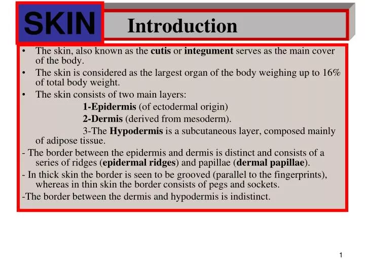
SKIN Introduction • The skin, also known as the cutis or integument serves as the main cover of the body. • The skin is considered as the largest organ of the body weighing up to 16% of total body weight. • The skin consists of two main layers: 1-Epidermis (of ectodermal origin) 2-Dermis (derived from mesoderm). 3-The Hypodermis is a subcutaneous layer, composed mainly of adipose tissue. - The border between the epidermis and dermis is distinct and consists of a series of ridges (epidermal ridges) and papillae (dermal papillae). - In thick skin the border is seen to be grooved (parallel to the fingerprints), whereas in thin skin the border consists of pegs and sockets. -The border between the dermis and hypodermis is indistinct.
THIN SKIN -(thumb) epidermis dermal papillae dermis sweat gland Pacinian corpuscle hypodermis subcutaneous BV fat
SKIN - EPIDERMIS, DERMIS AND HYODERMIS hairs cornified epithelium (stratified squamous) Epidermis dermal papillae Dermis (pegs) Meissner’s sweat gland corpuscle Hypodermis hair follicle subcutaneous fat Pacinian corpuscle
Functions of skin 1) Mechanical protection. The stratified keratinous epithelium of the skin provides mechanical protection against external abrasions or injury and against invasion of foreign objects. 2) Thermoregulation. plays a major role in regulation of body temperature, in particular by means of arterio - venous anastomoses. 3) Osmoregulation. regulation of body fluids and ions, and also protects against fluid loss. 4) Excretion and secretion. possesses exocrine glands (sweat glands, sebaceous glands). 5) Sensory reception. The skin receives sensory stimuli from the external environment. Several different types of receptor present in the skin respond to a variety of stimuli such as touch, pressure, heat, cold, pain. 6) Metabolic functions. The prohormone of vitamin D is present in the skin and is stimulated by ultraviolet light to provide the initial stage of the pathway leading to the synthesis of vitamin D metabolites. 7) Absorption. many drugs, can be absorbed through the skin (transdermal absorption). Encapsulation of drugs in a skin patch and their slow delivery can result in a continuous and steady absorption. Examples: 1-nitroglycerine (for cardiac therapy), 2- nicotine (to help addicted cigarette smokers quit), 3- estrogen (for postmenopausal replacement) or drugs to counter motion sickness. The skin is a very mobile tissue and needs the capability to stretch in areas of the body that are not static. This is well illustrated by considering the skin covering the elbow.
SKIN APPENDAGES - Skin appendages (derived from epithelium) include: 1-hair 2-nails 3-exocrine glands (sweat glands, sebaceous glands) • The free surface of the skin is not smooth, but has a series of fine grooves. In areas of the skin lacking hair these are more distinct. In particular the grooves are most pronounced on the thick skin of the hands (fingerprints) or the feet. • The skin in adults covers a total surface area of about 2.2m2. The surface area increases approximately sevenfold during the lifetime from an infant to adult and it is important to estimate skin surface in order to calculate dosage of drugs to be administered. The surface : volume ratio in young children is much greater than that of adults and consequently their control of body temperature is more problematic. A child loses heat more rapidly than an adult. • The thickness of the epidermis varies from 0.12-0.17 mm over most surfaces of the body ("thin skin"), but may reach a thickness of 0.8 mm on the palms of the hands and up to 1.4mm on the soles of the feet ("thick skin"). The terms thin skin and thick skin refer to the epidermis only and not to the total thickness of the skin. • Thick skin (of the palms and soles) is hairless, lacks pigment and is sometimes described as glabrous. Thin skin, in contrast, has hair, sebaceous glands and is pigmented.
The Epidermis • The epidermis consists of stratified squamous epithelium with a layer of keratin. The development of this layer of keratin was very important in evolution as it allowed the emergence of vertebrates from a fluid environment to a terrestrial environment. - 4 cell types, with different origins, are distinguished in the epidermis: 1- Keratinocytes (keratin production) 2- Melanocytes (pigment production) 3- Langerhans cells (immune system) 4- Merkel cells (diffuse neuroendocrine system).
The Keratin System • Keratinocytes (derived from ectoderm) are the dominant cell type of the epidermis, and are part of the keratinizing system, involved in the cornification or keratinization of the skin. • The most superficial cells are essentially dead cells, or scales, primarily composed of keratin. • These superficial keratinized cells are continuously being lost (desquamation or exfoliation) and need to be replaced. • New keratinocytes are continuously formed in the basal layers of the epithelium and mitotic figures are commonly seen in these layers. • The process of differentiation of the basal cells into the desquamating superficial scales is known as cytomorphosis and takes about 15 days in infants and up to 30 days in adults. • The epidermal layer is therefore being totally replaced in a relatively short period. • The skin disorder, psoriasis, is characterized by a very rapid turnover of keratinocytes (7 days). • The morphological changes occuring in the keratinocytes and the layers are best seen in thick skin.
Epidermis of thick skin • A- Basal layer (Stratum basalis) • B- Spiny layer (Stratum spinosum) • C- Granular layer (Stratum granulosum) • D- Clear layer (Stratum lucidum) • E- Horny layer (Stratum corneum)
EPIDERMIS STRATUM CORNEUM STRATUM GRANULOSUM STRATUM SPINOSUM VASCULAR PAPILLA STRATUM GERMANITIVUM
A- Basal layer (Stratum basalis) • The epithelial cells of the basal layer are closest to the basal lamina and to the underlying dermis. • These cells are typically columnar or cuboidal with many examples of mitosis. • At the ultrastructural level the most basal cells have unique structures on their basal plasmalemma known as hemidesmosomes (like half a desmosome). • The cytoplasm of the cells has tonofibrils (visible by light microscopy), which are seen to be tonofilaments (7-8 nm diameter) at the ultrastructural level. These tonofilaments are characterized as intermediate filaments composed of prekeratins. • As the basal cells continue to develop and move into the more superficial levels, the tonofilaments can constitute up to 50% of the proteins of the cells.
B -Spiny layer (Stratum spinosum) • The keratinocytes of the spiny layer are more rounded or oval and at the light microscope level there appear to be pronounced gaps between the cells. • The cells appear to be connected by "spines". • These spines, seen by ultrastructure, are composed of prominent desmosomes. • Tonofibrils are very dominant in these cells. • Mitoses are also common in the spiny layer. • The basal layer and the spiny layer together are known as the Malpighian layer or germinative layer (as this is the area of mitoses and generation of new keratinocytes).
STRATUM SPINOSUM PRICKLE CELLS TEM- DESMOSOME desmosomes PRICKLE CELLS IN STRATUM SPINOSUM TONOFIBRILS
C - Granular layer (Stratum granulosum) • This layer consists of 2-5 rows of fairly flattened cells characterized by large (1-5m) cytoplasmic basophilic granules known as keratohyalin granules. • Keratohyalin consists of phosphorylated histidine -rich proteins and cystine - containing proteins and is the precursor of keratin. • These granular cells also membrane-coating granules, which secrete their lipid-containing contents by exocytosis on to the surface of the cells. • This secretion provides the water impermeable barrier of the skin.
D - Clear layer (Stratum lucidum) • This is seen as a thin, undulating layer, stained well with eosin. • The nuclei of the cells are not well distinguished. • The cytoplasm of the cells contains a substance called eleidin, apparently derived from keratohyalin. • Relatively little is known about eleidin or its significance.
E - Horny layer (Stratum corneum) • The cells of the horny layer are flattened and their nuclei can no longer be distinguished. • The cells show a thickening of their plasmalemma (up to 15 nm). • The cells are packed with keratin. • Keratin belongs to the scleroproteins. • The keratin molecule is rich in disulfide bonds and is anisotropic (birefringent) when examined by polarizing microscopy.
EPIDERMIS sweat pore stratum disjunctum stratum corneum stratum lucidem stratum granulosum melanocyte stratum spinosum papillary layer of dermis stratum germanitivum sweat gland sensory vascular (conducting) papilla dermis papilla reticular layer sweat gland of dermis (secretory)
Epidermis of thin skin • The epidermis of thin skin is much thinner and less well developed than that of thick skin. • The basal layers and horny layers are always present, but much thinner, however, the clear layer is usually not found and the granular layer is very thin, consisting of a single or double row of cells. • Skin cancer derived from basal layers is known as basal cell carcinoma, whereas skin cancer developing from more superficial cells is known as squamous cell carcinoma. • These skin cancers are extremely common and constitute up to 30% of all the tumors of the body. • They are usually not life-threatening and relatively easy to treat. THIN SKIN - ABDOMEN
The Pigmentary System • The color of the skin depends on its thickness, and the degree of underlying vascularization, especially the oxyhaemoglobin. • If blood vessels are contracted, such as in cold environments, the skin is more pallid, whereas if we exert ourselves the vessels dilate and we appear redder. • The yellowish color of skin is due to the pigment carotene (excessive dietary intake of carotene-containing foods may induce a more yellow or orange coloration). • Skin color can sometimes provide diagnostic clues to underlying disorders (anemia, cyanosis, hepatitis).
The main pigment of the skin, which provides the main component of skin coloration, is melanin. • Melanin is produced by melanocytes, derived from the embryonic neural crest. • Melanocytes are found in the more basal layers of the epidermis. • They are rounded cells with processes that extend between the adjacent keratinocytes. • They are not connected to the keratinocytes by any structures and lack desmosomes. • The melanocytes need to synthesize the enzyme, tyrosinase, in order to synthesize melanin. • Albinos have an inborn error of metabolism and are unable to synthesize tyrosinase and are consequently unable to produce melanin. • Melanin is lacking in thick skin. • The palms of the hands and the soles of the feet are unpigmented, even in dark-skinned people. • The synthetic process of melanin formation within the melanocytes involves a simple biosynthetic pathway :
Tyrosine is converted (by tyrosinase) to DOPA (dihydroxyphenyl alanine) and DOPA- quinone, which is converted to the dark pigment melanin. • The melanin is seen in histological sections as a dark brown color in both melanocytes and adjacent keratinocytes. • The melanin is transferred to the keratinocytes (apparently by phagocytosis) and as a result more melanin may accumulate in the keratinocytes than in the melanocytes. • The development of melanin granule formation at the ultrastructural level reveals rugby-ball shaped bodies formed by the Golgi bodies. • These initially have little pigment and are known as premelanosomes, but soon become packed with melanin and are known as melanosomes. • It is the melanosomes (melanin granules) that are taken up by the keratinocytes.
The number of melanocytes is fairly constant in all races (about 1000/mm2 on most of the body, but 2000/mm2 on the scrotal skin). The difference between races is in the rate of expression of melanin formation, being much greater in people with dark skins. • Skin pigmentation is genetically determined, though in lighter-skinned people pigmentary changes commonly result from : -(a) endocrine disturbances (such as excess ACTH) -(b) pregnancy (including changes in the color of the areola of the nipples) -(c) exposure to ultraviolet light ("suntan"). • The tanning process following exposure to sunlight is a protective measure to reduce the damage of ultraviolet light to the underlying tissues. • Following initial inflammatory changes, there is increased synthesis of tyrosinase and melanin. • Ultraviolet light is also a cause of age-related skin changes, including skin thickening, hardening and increased wrinkling. • Malignant melanoma is a serious form of skin cancer that develops from proliferation of melanocytes. • Excess ultraviolet light exposure appears to be a predisposing factor in the development of melanoma.
SKIN PIGMENTATION AND MELANIN PIGEMENTED SKIN Malphigian Cell desmosome melanocyte melanosomes Pigment in Malphigian cells hemi- desmosome stratum germanitivum basement membrane
Langerhans cells • Langerhans cells are found in the epidermis and number 400-1000 per mm2. • They are star-shaped cells found mainly in the spiny layer and can be demonstrated by impregnation with gold chloride techniques. • They function as antigen-presenting cells of the immunesystem of the skin. • At the ultrastructural level their cytoplasm is seen to possess characteristic rod-like granules known as Birbeck's granules.
Merkel cells • These are found in thick skin and at the ultrastructural level are seen to possess numerous cytoplasmic granules, typical of polypeptide-secreting endocrine cells. • These cells are commonly associated with free nerve endings. • The function of Merkel cells is still not fully established, though it is thought they may function as sensory mechanoreceptors or produce local neuroendocrine secretions. • It is believed that they belong to the diffuse neuroendocrine system of the body.
The Dermis • The dermis is the layer of connective tissue beneath the epidermis and is derived from mesoderm. • It is difficult to measure the thickness of the dermis as the border with the underlying hypodermis is not distinct. • Over most of the body the dermis is 1-2 mm thick, though in thick skin it may reach 3 mm or more. • The dermis of the dorsal side of the body is in general thicker than that of the ventral side. • Usually the dermis is thinner in females than in males. • Two distinct regions are present in the dermis A-papillary layer B-reticular layer
The papillary layer • includes the area of the dermal papilla and consists of loose connective tissue. • The main blood vessels that supply the epidermis are located in the papilla. (As the epidermis is avascular, the location of the blood vessels in the papillae reduces the distance needed for diffusion). • The dermis is well-vascularized and has many arterio -venous anastomoses, which play important roles in thermoregulation of the body. • Up to 4.5% of blood volume is found in the dermis. • In cold external environments the body needs to conserve heat and the blood can go directly from the arteries to the veins, by-passing the capillary bed. • In hot environments, when we need to cool the body more, the blood is allowed to pass into the capillary beds.
The reticular layer • is thicker than the papillary layer and consists of a more dense, irregular connective tissue composed mainly of collagen bundles. • The term reticular refers to the netlike arrangement of these bundles. • Both the papillary and the reticular layers have elastic fibers, responsible for the elasticity and flexibility of the skin. • With aging the collagen fibers thicken and cross-link and the elastic fibers lose much of their elasticity, causing increased wrinkling of the skin. • The process of age-related wrinkling is increased in cigarette smokers and in people exposed excessively to ultraviolet light. • Sun exposure is a major factor in age-related damage to skin.
The dermis of the face is the site of insertion of the muscles of facial expression. • These striated muscles are vestiges of a more extensive subcutaneous muscle layer found in many mammals (Panniculus carnosus), which is used to shake wet fur dry, or dislodge insects. • Several epithelial derivatives are also found in the dermis (hair, sweat glands, sebaceous glands). • Several types of sensory receptors are also found in the dermis. • Meissner's corpuscles are tactile mechanoreceptors found in dermal papilla, especially in thick skin, lips, eyelids, genitalia, nipples. • Pacinian corpuscles ( Vater – Pacini ) are mechanoreceptors common in the dermis of thick skin.
The Hypodermis • The hypodermis consists of loose connective tissue and is composed mainly of adipose tissue.
Hairs • Hairs are one of the unique characteristics of mammals. • They are thin filaments of keratin that develop in the dermis from epithelial invaginations of the epidermis, which interact with a germinative center of the dermis - the dermal papilla. • Hairs are found on skin of most parts of the body, apart from thick skin (palms of the hands, soles of the feet), lips, glans penis, labia minora and clitoris. • The number of hairs on the face are about 600 hairs per cm2, whereas in the other parts of the body they are about 60 per cm2. • The appearance of hairs (length, thickness, pigmentation, density) depends on many factors including: age, race, sex, hormones, location on the skin. • Hairs grow discontinuously, with periods of growth followed by periods of inactivity. • The hairs on the head may have a growing phase of 3 years or more, whereas in other areas of the body the growing phase may be only 3 months or so. • Hair growth is not synchronous and in any particular site, there will be hairs in active growth phases as well as hairs in inactive phases. • This is described as mosaic growth.
The development of hairs involves epithelial invaginations (proliferation and downgrowth) from the epidermis into the connective tissue of the dermis. • The tubular epidermal invagination is known as the hair follicle. • The most terminal portion of the invagination expands to form the hair bulb. • A connective tissue sheath surrounds the follicle (dermal sheath). • During development there is a process of induction during which a concentration of connective tissue (dermal papilla) becomes associated with and surrounded by the hair bulb. • The association of the dermal papilla and the hair bulb is essential for hair growth. • Blood vessels invade the dermal papilla in order to supply the necessary nutrients and hormones for hair growth. • During hair development two epithelial protrusions develop on the follicle. • The upper protrusion is the site for the development of the sebaceous gland. • The lower protrusion is the site for attachment of a smooth muscle bundle, known as the arrector pili. • The site of insertion of the arrector pili muscle is in the papillary layer of the dermis. • Contraction of the arrector pili muscles causes extrusion of the oily secretion (sebum) from the sebaceous glands, via a short duct onto the surface of the growing hair. • The arrector pili muscles contract in response to fear, anger or cold and are responsible for gooseflesh.
The germinative area of the hair is a short cone-like region just above the hair bulb. • This is the region where the hair cells become keratinized and pigmented. • When examined in more detail, the epithelial components of the hair are divided into an outer epithelial sheath, continuous with the Malphighian layer of the epidermis and an inner epithelial sheath, which extends only to the level of the sebaceous glands. • The epithelial cells of the outer epithelial sheath grow downwards in the direction of the bulb and continue upwards in the inner epithelial sheath. • The hairs have a central core (medulla) composed of large vacuolated cells, with relatively little pigment or keratin. • This is absent in the fine lanugo hairs of the newborn, that are usually shed within a month or so. • The medulla is surrounded by the cortex, composed of cells that are usually very pigmented and keratinized. • The outermost part of the hair is the cuticle, which is composed of overlapping scales of hard, transparent (non-pigmented) keratin.
Melanocytes, situated in the hair bulb, transfer their pigment to the cells of the medulla and cortex. With aging the melanocytes produce less melanin resulting in white hair. • The part of the hair that projects from the skin consists of non-viable (dead) cells. • The outer epithelial sheath lies on a thick basal lamina (the "glassy membrane"), which separates the follicle from the surrounding dermal sheath. • During periods of cessation of hair growth, the hair papilla becomes detached from the hair bulb and the hair follicle adopts a club-like appearance (club hair). • It is during these periods that hair loss is experienced. • Hair growth is influenced by endocrine factors and in particular the sex steroids. • The changes in hair distribution associated with sexual maturity are the result of changes in endocrine secretion and development of receptors to androgens in particular.
HAIR FOLLICLE hair epidermis external root arrector pili sheath muscle internal dermi root sebaceous basement sheath gland membrane hair shaft hair dermis epidermis papilla bulb dermal mitosis) papilla
HAIR FOLLICLE - TRANSVERSE SECTION Hair (cuticle, cortex, medulla) internal external root sheath root sheath dermis Huxley’s glassy C layer membrane M Henle’s layer Henle’s external glassy Huxley’s membrane root sheath dermis medulla cortex cuticle (keritinized cells)
HAIR FOLLICLES IN SCALP HAIR FOLLICLES – T/ SEC
Sebaceous glands • The sebaceous glands are dermal exocrine glands associated with hairs and which secrete an oily substance (sebum) on the growing hair. • The sebum is important in maintaining the flexibility of the hair. • The glands are composed of alveoli and a short secretory duct. • The outermost cells of the gland are filled with lipid droplets. • These cells are secreted in their entirety (holocrine secretion). • The extrusion of the sebum results from the contraction of the arrector pili muscles. • The sebum secretion is influenced by sex hormones (androgens and estrogens) and during hormonal imbalance during puberty may result in adolescent acne. • Sebaceous glands are not present in thick skin.
SEBACEOUS GLAND sebaceous gland disintigrating cells (holocrine secretion) hair shaft hair follicle sweat glands arrector pili muscle sebaceous gland (simple-branched alveolar
Sweat glands • simple exocrine glands that secrete sweat. • The secretory units are simple convoluted tubular epithelial structures in the dermis and a straight secretory duct. • The convolutions of the secretory unit are seen in histological preparations as many associated profiles of the same unit. • The secretory ducts are surrounded by myoepithelial cells, which on contraction cause the expulsion of the sweat. Two sorts of sweat glands are found: 1-Eccrine (Merocrine) sweat glands • found all over the body including the thick skin. • In thick skin sweat pores are conspicuous in the thick outer layer of keratin. • Fingerprints are formed by secretions derived from the sweat glands. • The eccrine sweat glands have cholinergic innervation.
2-Apocrine sweat glands • large sweat glands located in the axilla (armpit) and also in association with the external genitalia and anus. • They have very large secretory units and myoepithelial cells, in which the apical part of the secretory cells including their contents, are secreted into the lumen (apocrine secretion). • The breakdown of this secretion by bacteria is the cause of the typical smell of sweat from the armpits. • The apocrine sweat glands only become functional at puberty and are influenced by sex steroids. (Young children do not have a noticeable sweat smell.) • It is possible that the secretions of apocrine sweat glands contain pheromones (as sex chemoattractants). • The apocrine sweat glands have adrenergic innervation.
ECCRINE SWEAT GLAND SWEAT GLAND (simple coiled tubular) spiral duct SPIRAL DUCT EPIDERMAL PEG CONDUCTING PORTION conducting secretory portion portion SECRETORY PORTION
Nails • Nails are horny plates of keratin on the dorsal surface of the terminal phalanges of the hand and foot (fingers and toes). • The nail plate is composed of tough, hard, clear non-desquamating keratin, which sits on the nail bed, composed of a layer of epidermal cells. • Surrounding the nail is the nail groove. • The formation of the nail and its keratin occurs in the nail root. • This region shows many mitoses of the epithelial cells. • These germinative cells of the nail root are known as the dorsal and ventral matrix. • Part of the ventral matrix (seen as a whitish half-moon near the dorsal nail groove) is called the lunula. • The keratin above the nail groove is thickened (eponychium). • Below the nail, near its free surface, the keratin is also thickened (hyponychium). • If the terminal portion of a finger is cut transversely, the lateral nail grooves are easily seen, and the clear nail plate is also obvious. • The nail bed, however, is seen to have many small undulations, in which small blood vessels and nerves are located.
NAILS (fingers and toes) Nail (dead, cornified eponychium epithelial cells) hyponychium nail bed nail root cuticle lunula eponychium nail hyponychium nail bed nail root
Meissner’s corpuscles - H & E Nerve endings in the skin • The skin has many sensory elements that respond to external impulses and signals. 1- Free nerve endings are non-encapsulated nerve endings in the epidermis and which respond to pain. 2- Meissner corpuscles are mechanoreceptors present in the dermal papilla. 3- Merkel corpuscles are mechanoreceptors surrounding hair follicles responsive to touch. 4- Pacinian corpuscles (Vater-Pacini) are found in the dermis of thick skin of fingers and respond to pressure and vibration. 5- Krause end bulbs are found in the dermis and respond to cold. sensory papilla Meissner’s corpuscle
Split skin grafts • In cases of trauma or burns in which the skin is damaged, skin transplants (autografts) can be taken to cover the wounds. • In these cases a thin slice of skin is removed (epidermis and dermis) from an uninjured region and transplanted to cover the lesion. • The epithelium of the skin rapidly proliferates to cover the wound and the epithelial components (hairs and glands) also rapidly recover. • The area from where the graft was taken also rapidly recovers. • The epidermal derivatives (hair and glands) rapidly grow outwards to form new epidermis.