Download

1 / 34
360 likes | 704 Vues
Chapter 1 Introduction to rheumatoid arthritis (RA). BR 8662 – 03/2009. Content. Rheumatoid arthritis (RA) Burden of RA RA etiology RA pathogenesis Systemic and extra-articular features of RA Articular effects of RA RA differential diagnosis RA diagnosis RA remission. Burden of RA.

E N D
Chapter 1Introduction to rheumatoid arthritis (RA) BR 8662 – 03/2009
Content • Rheumatoid arthritis (RA) • Burden of RA • RA etiology • RA pathogenesis • Systemic and extra-articular features of RA • Articular effects of RA • RA differential diagnosis • RA diagnosis • RA remission
Burden of RA • An autoimmune, chronic, destructive inflammatory disease with both articular and systemic effects1 • Impairs health-related quality of life (QOL)2 • Cause of disability and mortality1 • Represents a major economic burden, for both individuals and society2 • Most common chronic autoimmune disorder3,4 • Global prevalence: about 0.5 – 1.0% worldwide • Prevalence and incidence of RA increase with age (peak at 70 and then decline) • Prevalence and incidence higher in women: 2 fold increase risk • Belgian prevalence of RA in population > 35 yr estimated at 0.914% (= mean European prevalence)5 • estimated number of patients > 35 yr in Belgium affected by RA is 57.473 1Smolen J and Aletaha D. Eur J Health Econ 2008;8(Suppl 2):S39-S47 2Lundkvist J, Kastäng F and Kobelt G. Eur J Health Econ 2008;8(Suppl 2):S49-S60 3Orozco G, Rueda B and Martin J. Biomed Pharmacother 2006;60:656-662 4http://www.who.int/healthinfo/statistics/bod_rheumatoidarthritis.pdf 5http//decisionresources/stellent/groups/public: 2005 estimation of prevalence and incidence of RA with mean European prevalence of 0.914%
RA etiology • Gender1: ♀ > ♂ • Genetic factors2: HLA-DRB1, PTPN22 • Environmental factors3-6: smoking, coffee intake, diet, obesity, previous blood transfusions and other autoimmune diseases • Biomarkers7 • Rheumatoid factor (RF) • Anti-cyclic citrullinated peptide antibodies (anti-CCP) • C-reactive protein(CRP) • Erythrocyte sedimentation rate (ESR) HLA = human leukocyte antigen 1http://www.who.int/healthinfo/statistics/bod_rheumatoidarthritis.pdf 2Orozco G, Rueda B and Martin J. Biomed Pharmacother 2006;60:656-662 3Symmons DP, et al. Arthritis Rheum 1997;40(11):1955-61 4Mikuls TR, et al. Arthritis Rheum 2002;46(1):83-91 5Pedersen M, et al. J Rheumatol 2005;32:1249-1252 6Silman AJ and Pearson JE. Arthritis Rheum 2002;4(Suppl 3):S265-S272 7Shovman O, et al. Clin Dev Immunol 2005;12(3):197-202
RA pathogenesis RA is an immune-mediated syndrome with participation of both the innate and the adaptive arm of the immune system Involvement of numerous cells of the immune system Dendritic cells T cells B cells Involvement of broad spectrum of cytokines Amplify inflammatory pathways Promote activation of non-immune cells as fibroblasts, chondrocytes and osteoclasts Ag=antigen; APC=antigen-presenting cell; DC=dendritic cell; IFN=interferon; IL=interleukin; MHC=major histocompatibility complex; MMP=matrix metalloproteinase; RANK=receptor activator of nuclear factor-κB; RANKL=RANK ligand; TCR=T-cell receptor; TLR=Toll-like receptor; TNF=tumor necrosis factor Scheinecker C, Smolen JS and Weynand CM, 2008
RA pathogenesis: The role of dendritic cells • Potent role in innate immune responses • Antigen-presenting capacity • Unique ability to activate naive T cells • Location: • in diverse tissues with high potential for invasion by pathogens • Epidermis, gastrointestinal mucosa • described in the synovial membrane in RA Scheinecker C, Smolen JS and Weynand CM, 2008
RA pathogenesis: The role of T cells in RA Macrophage (Auto) antigen TNF, IL-12, IL-1, IFN1, IL-2 Dendritic cell T cell IFN- IL-2 IFN- CD80/CD86 IL-17 TH1 CD28/CTLA-4 IL-15 TCR MHC II IL-18, IL-32 TNF, IL-6, IL-1 TH1=T-helper 1 cell; CTLA-4=cytotoxic T-lymphocyte antigen 4 Smolen JS et al. Lancet 2007;370:1861-1874; Scheinecker C, Smolen JS and Weynand CM. 2008
RA pathogenesis: The role of B cells in RA TH1 B cell autoantibodies (RF and others) receptor for Fc portion of IgG CD4 CD28/CTLA-4 CD80/CD86 CD40 CD40L TCR MHC II (Auto)antigen Complement receptor Co-stimulation T cell help IC formation Plasma cell Macrophage Complement TNF, IL-6, IL-1 IC=immune complex Smolen JS, et al. Lancet 2007;370:1861-1874; Scheinecker C, Smolen JS and Weynand CM. 2008
pro-inflammatory cytokines anti-inflammatory cytokines RA pathogenesis: Cytokines and RA • Broad spectrum of cytokines is involved in the RA pathogenesis • joints of early-stage RA • Th2 cytokines such as IL-4 • more established RA • Th1 cytokines such as IL-2, IFN-γ, TNF-α, granulocyte-macrophage colony-stimulating factor (GM-CSF) • expression of multiple pro-inflammatory cytokines deriving from cells in the inflamed synovium such as macrophage-like type A synoviocytes Scheinecker C, Smolen JS and Weynand CM. 2008 Firestein GS. Nature 2003;423:356-361
RA pathogenesis: Pro-inflammatory cytokines and RA • Pro-inflammatory cytokines play a significant role in promoting joint as well as systemic inflammation in RA • TNF-α, IL-1, IL-6 • Act on various cell populations • Partially stimulate each other • Major role in bone destruction and loss of cartilage • Partly responsible for peri-articular osteoporosis Ab=antibodies; Ch=chondrocyte; EC=endothelial cell; Fb=fibroblast; Hep=hepatocyte; HSC=hematopoietic stem cell; KC=keratinocyte; MK=megakaryocyte; MMP=matrix metalloproteinases; OC=osteoclast Scheinecker C, Smolen JS and Weynand CM. 2008
RA pathogenesis: The role of fibroblasts and chondrocytes in cartilage damage during RA TNF, IL-1, IL-6 Synovial fibroblast MMPs Chondrocyte Cartilage Bone MMP=matrix metalloproteinases Smolen JS et al. Lancet 2007;370:1861-1874
RA pathogenesis: Osteoclasts have a central role in mediating bone destruction in RA IL-6, TNF-, IL-1 Indirect Direct RANKL expression Synovial fibroblast Activation RANKL RANK M-CSF Differentiation Osteoclast Macrophage/Monocyte Cathepsin K Bone M-CSF = macrophage colony stimulating factor; RANKL = receptor activator of nuclear factor κβ ligand Scheinecker C, Smolen JS and Weynand CM. 2008; Schett G. Arthritis Res Ther 2007;9:203 (doi:10.1186/ar2110) Smolen JS, et al. Lancet 2007;370:1861-1874; Teitelbaum SL, et al. Science 2000; 289:1504–1508; Kudo O, et al. Bone 2003; 32:1–7.
RA pathogenesis: The role of VEGF in pannus formation Angiogenesis in RA is a key process in formation and maintenance of the pannus Expansion of the synovial lining of joints in RA Subsequent invasion by the pannus of underlying cartilage and bone Necessity of increased vascular supply to synovium, to cope with the increased requirement for oxygen and nutrients Vascular endothelial growth factor (VEGF) has been identified as a key element in the development of new blood vessels Synovial fibroblasts VEGF Pannus formation Endothelial cells Paleolog EM. Arthritis Res 2002;4(Suppl 3):S81–90
RA pathogenesis: Up-regulation of adhesion molecules facilitates cellular infiltration in RA In RA, endothelial cells within the synovial membrane are activated and express cellular adhesion molecules (CAMs) that promote the recruitment of inflammatory cells into the joint The ability of cells to adhere to other cells and extracellular matrix through CAMs plays a critical regulatory role in synovial lining hyperplasia Leucocyte-endothelial cell interaction Local activation of endothelial cells and leukocytes by chemokines leads to rapid upregulation of CAMs which facilitates firm adhesion and trans-endothelial migration of leukocytes from the circulation to the into tissues Interaction of leukocytes with synovial endothelial cells is a critical step in the development of synovial inflammation Synovial sublining Infiltration by T and B lymphocytes, dendritic cells and macrophages High degree of organization of complex lymphoid microstructures Synovial lining Two cell types: fibroblast-like synoviocyte (FLS) and macrophage-like synoviocyte (MLS) Synovial lining becomes hyperplastic with increased numbers and activation of both FLS and MLS Extension into the joint space with attachment to cartilage and invasion and degradation CAMS include selectins, integrins, the immunoglobulin superfamily and cadherins Agarwal SK and Brenner MB. Curr Opin Rheumatol 2006;18:268–276
RA pathogenesis: Chemokines and leukocyte recruitment in RA Leukocyte infiltration into the joint space and tissues is an essential component of the pathogenesis of RA Chemokines and their receptors play a key regulatory role in organ-specific leukocyte trafficking and activation by affecting integrin activation, chemotaxis, effector cell function and cell survival Chemokines are small peptide molecules Secreted from a wide variety of cells Involved in the innate and adaptive immune response Constitutively expressed or induced during inflammation Cells that express the appropriate chemokine receptor migrate across the vessel wall towards the source of the chemokine Tarrant TK and Patel DD. Pathophysiology 2006;13:1–14
Anaemia1 Osteoporosis2,7 Pain3 Fatigue4,7 Interstitial lung disease5 Pleuritis5 Pericarditis5 Vasculitis5 Cachexia6 Autoantibody production7 Fever7 Hypergammaglobulinaemia7 Thrombocytosis7,8 Increased cardiovascular disease9 Systemic and extra-articular features of RA 1Fitzsimons EJ, et al. Lancet 2002;360:1713–1714 2Solomon DH, et al. Arthritis Rheum 2006;55:873–8773McCabe CS. Musculoskeletal Care 2004;2:75–89 4Lundkvist J, Kastäng F and Kobelt G. Eur J Health Econ 2008;8(Suppl 2):S49-S605Turesson C and Matteson EL. Curr Opin Rheum 2004;16:206–2116Walsmith J and Roubenoff R. Int J Cardiol 2002;85:89–997Yoshizaki K, et al. Springer Semin Immunopathol 1998;20:247–2598Kacena MA and Horowitz MC. Curr Opin Rheum 2006;18:405–410 9Snow MH and Mikuls TR. Curr Opin Rheum 2005;17:234–241
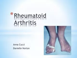
Articular effects of RA • Affects small joints of the hands and feet, as well as large joints • Symmetric character, frequently affecting the same joint regions on both sides • Joint destruction is the major hallmark of RA and is usually progressive • Inflammation/proliferation of the synovium with pannus formation • Bone erosions and cartilage degradation lead to structural joint damage with joint space narrowing • Destruction of supporting articular structures • A major cause of disability Smolen J and Aletaha D. Eur J Health Econ 2008;8(Suppl 2):S39-S47; Firestein GS. Nature 2003;423:356–361 Scheinecker C, Smolen JS and Weynand CM, 2008
Joint destruction is the hallmark of RA T cell B cell Plasma cell Synovial villi Angiogenesis Neutrophils Pannus Eroded bone Healthy joint Rheumatoid joint Femur Capsule Cartilage Synovial membrane Tibia Synoviocytes Choy EHS and Panayi GS. N Eng J Med 2001;344:907–916
SLE High-titer ANA Anti-ds-DNA Low serum complement Nephritis Polymyalgia rheumatica Elderly patients Peripheral synovitis Prominent proximal muscle stiffness Very high ESR Gout Characteristic gouty erosions Crystals in synovial fluid Spondyloarthopathies Lack of symmetry Distribution of affected joints Absence RF Skin lesions(e.g. psoriatic arthritis) Osteoarthritis Morning stiffness uncommon Involvement of wrist and MCP joints uncommon RF negative Non-inflammatory synovial effusions RA: differential diagnosis MCP=metacarpophalangeal Firestein GS. APC Medicine. WebMD;2006. Rheumatology:II Rheumatoid arthritis.
RA: diagnosis • Clinical features • Imaging • Laboratory evaluation • Disease scoring systems • Disease activity: ACR, DAS, EULAR, SDAI and CDAI • Quality of life scores: HAQ, SF-36, MOS sleep score, FACIT-fatigue • Radiographic scores: Larsen score, Sharp score, Sharp/van der Heijde score (SHS) and Genant modified Sharp score
RA: Clinical features • Most common: Insidious onset followed by progression to polyarticular inflammation • Abrupt and acute possible • Symmetrical involvement • Small joints of hands and feet at the outset, but also large joints • Morning stiffness > 30 minutes (often up to several hours) • Swelling, warmth, tenderness to palpation • Limitation of active and passive range of motion Firestein GS. APC Medicine. WebMD;2006. Rheumatology:II Rheumatoid arthritis.
Imaging in RA: Conventional radiography • X-ray is the traditional gold standard for assessment of joint damage in RA1,2 • Most frequently hands and feet • Joints in these sites are affected in most RA patients • Changes in these small joints correlate with abnormalities in large joints • Focus on bone erosions and joint space narrowing • 30% of patients have bony erosions at time of diagnosis (>60% at 2 years)3 • Validated scoring methods available • Sharp score • Genant-modified Sharp score • Sharp/van der Heijde score (SHS) • Advantages • Low cost, high availability, standardization and blinded centralized reading, reproducibility, helpful in differentiation from other joint conditions • Disadvantages • Superimposition, ionizing radiation, insensitivity to early bone damage, insufficiency for assessment of soft tissue changes 1Østergaard M. Best Pract Res Clin Rheumatol 2005;19(1):91-116 2van der Heijde D, et al. Rheumatology 1999;38:941-947 3O'Dell JR. N Engl J Med 2004;350:2591-2602
Imaging in RA: US and MRI • Ultrasound (US): • Advantages: • Non-invasive, relative inexpensive, absence of harmful radiation, rapidity of imaging, easy repeatable, examination of several joint regions at one session, potential for being performed by rheumatologists, visualization of both inflammatory and destructive disease manifestations and superior to X-ray in detecting bone erosions early in RA • Disadvantages • Not all areas are accessible, skilled operator • Magnetic Resonance Imaging (MRI) • Advantages: • Allows assessment of all the structures involved in RA and is more sensitive than clinical examination and X-ray for detection of inflammatory and destructive joint changes in early RA; no ionizing radiation • Disadvantages: • Higher cost, lower availability, longer examination times, only few joints per session Østergaard M. Best Pract Res Clin Rheumatol 2005;19(1):91-116
RA: Laboratory evaluation • Elevation of common non-specific serum markers of inflammation contributes to the assessment of disease activity level1 • Erythrocyte sedimentation rate (ESR) • C-reactive protein (CRP) • Rheumatoid factor (RF)1 • Usually appears in the first year of the disease • ~80% of RA patients are RF+ • Associated with more rapidly evolving the more erosive disease and a higher incidence of extra-articular manifestations2 • 1% to 5% of healthy persons test positive for RF • RF also associated with other chronic diseases • Anti-cyclic citrullinated peptide (anti-CCP)1,3 • Highly specific (specificity ~ 90%) • The level of anti-CCP may predict the development of more serious disease and increased risk for joint damage • Antinuclear antibody (ANA)2 • Positive in 20–30% of RA patients • More common in patients with extra-articular manifestations 1Firestein GS. APC Medicine. WebMD;2006. Rheumatology:II Rheumatoid arthritis; 2Matsumoti AK. http://www.hopkins-arthritis.org/arthritis-info/rheumatoid-arthritis/rheum_clin_pres.html3Vencovský J, et al. Ann Rheum Dis 2003;62:427-430
American College of Rheumatology (ACR) criteria for the classification of RA • The American College of Rheumatology revised the criteria for classification of RA in 1987 • A patient has RA if he/she has satisfied at least 4 of these 7 criteria with criteria 1-4 present for at least 6 weeks • Morning stiffness in/around joints of ≥ 1 hour before maximal improvement • Arthritis with soft-tissue swelling or fluid in ≥ 3 of 14 joints/joint groups • Arthritis of hand joints with ≥ 1 area swollen in wrist, MCP or PIP • Symmetric arthritis • Rheumatoid nodules subcutaneous in specific places • Serum rheumatoid factor at a level above the 95th percentile • Radiographic changes suggestive of joint erosion ACR was formerly knwon as the American Rheumatism Association Arnett FC, et al. Arthritis Rheum 1988;31(3):315-324
ACR response measure • Evaluation of change in clinical status over time • ACR 20%, 50% and 70% improvement criteria (ACR20/50/70) • Restricted to analysis of clinical trials and not useful in clinical management of the individual patient Requirements(i.e. ACR20 response) Measure of disease activity (ACR core set) 20% improvement 20% improvement Tender joint count Swollen joint count Patient pain assessment (VAS) Patient global assessment of disease activity (VAS) Physician global assessment of disease activity (VAS) Patient assessment of physical function (HAQ) Acute-phase reactant (ESR or CRP) 20% improvement in 3 of the 5 measures Felson DT, et al. Arthritis Rheum 1995;38:727-735 Ranganath VK, Khanna D and Paulus HE. Clin Exp Rheumatol 2006;24(Suppl 43):S14-S21
Disease Activity Score: DAS28 • Evaluation of disease activity at a specific timepoint • Information about 4 disease variables is needed for DAS score calculation • Swollen joint count using 28-joint counts (SJC) • Tender joint count using 28-joint counts (TJC) • Acute-phase reactant (ESR or CRP) • Patients general health (GH) or global disease activity measured on a Visual Analogue Scale (VAS) Tender joints/swollen joints DAS28 = 0.56 × (TJC28) + 0.28 × (SJC28) + 0.70 × ln(ESR) + 0.014 × GH ESR Patients assessment of general health Prevoo MLL, et al. Arthritis Rheum 1995;38(1):44-48 http://www.das-score.nl
DAS28 reference values Remission • DAS28 is a sensitive discriminator between patients with high and low disease activity and between active and placebo tretaed patient groups • Serial measurements of DAS28 are strong predictors of physical disability and radiological progression • DAS28 provides a number on a scale from 0 to 10 indicating the current activity of RA 2.6 Low disease activity 3.2 Intermediate disease activity 5.1 High disease activity http://www.das-score.nl
European League Against Rheumatism response criteria for RA • EULAR response criteria are based on the DAS • Improvement in the DAS from baseline • DAS attained during follow-up DAS28 at endpoint Improvement in DAS28 from baseline 0.6 >1.2 >0.6 and 1.2 Good 3.2 Moderate >3.2 and 5.1 None >5.1 van Gestel AM, et al. Arthritis Rheum 1996;39(1):34-40 EULAR handbook of clinical assessments in rheumatoid arthritis. 2004
Other composite indices in RA: SDAI, CDAI SDAI: Simplified Disease Activity Index; CDAI: Clinical Disease Activity Index Aletaha D and Smolen J. Clin Exp Rheumatol 2005;23(Suppl 39):S100-S108
Health-related quality of life (HR-QOL) assessments in RA • Health Assessment Questionnaire Disability Index (HAQ-DI)1,2: • Assessment of functional disability • Scale of 20 activities of daily living (ADL) in 8 categories • Dressing, arising, eating, walking, bathing, reaching, gripping, performing errands • Scaled from 0 (without any difficulty) to 3 (unable to do) • Total HAQ score = mean score derived from 8 scores + VAS assessment of pain and global status • Modifications of HAQ have been developed for ease of scoring in standard clinical care • Multidimensional HAQ (MDHAQ) • 13 items: 10 concerning physical function and 3 concerning sleep, anxiety and depression • VAS for pain, global status and fatigue, length of morning stiffness and change in status • Short-Form 36 (SF-36)1: • Evaluation of health status • 8 scales: physical functioning, role limitations due to physical health, bodily pain, general health perceptions, vitality, social functioning, role limitations due to emotional health and mental health • 2 summary scores for physical and mental components • Medical Outcomes Study (MOS) sleep scale3: • 12-Item self-report measuring specific aspects of sleep • Functional Assessment of Chronic Illness Therapy-Fatigue scale (FACIT-Fatigue)4 1Russell A. Pharmacoeconomics 2008;26(10):831-846 2Pincus T. Bull NYU Hosp Jt Dis 2006;64(1-2):32-39 3Hays RD and Stewart AL. Durham NC, Duke University Press 1992;235-259 4http://www.facit.org
Radiographic scores in RA • Larsen method and Sharp method1 • Includes erosion scores (ERO) and joint space narrowing (JSN) • Sharp/van der Heijde score (SHS)2 • ERO scores: 0 to 5 in hands and 0 to 10 in feet with highest score indicating total destruction • JSN score from 0 (normal) to 4 (ankylosis) • Total score is the sum of scores for erosions and JSN with a maximum of 448 • Genant-modified Sharp score3 • ERO and SJN in both hands 1Sokka T. Bull NYU Hosp Jt Dis 2008;66(2):166-168 2van der Heijde D, et al. Rheumatology 1999;38:941-947 3Genant HK, et al. Arthritis Rheum 1998;41(9):1583-1590
Durable clinical remission is an important goal in management of RA Remission in RA refers to a near complete suppression of disease activity or an absence of discernable disease activity Clinical remission does not connote an absence of disease progression Radiologic progression among RA patients in remission Defining remission requires objective outcome measures ACR complete clinical remission: ≥5 criteria lasting ≥ 2 consecutive months Morning stiffness <15 minutes No fatigue No joint pain by history No joint tenderness or pain in motion No soft-tissue swelling in joints or tendon sheats ESR <30 mm/hr (female) or < 20 mm/hr (male) DAS28 remission: DAS28<2.6 FDA major clinical response: ≥ACR70 for ≥6 continuous months RA: Remission as ultimate goal Sesin CA and Bingham CO. Semin Arthritis Rheum 2005;35:185-196 Molenaar ETH, et al. Arthritis Rheum 2004;50:36-42