Download
1 / 46
510 likes | 885 Vues
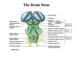
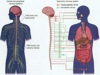
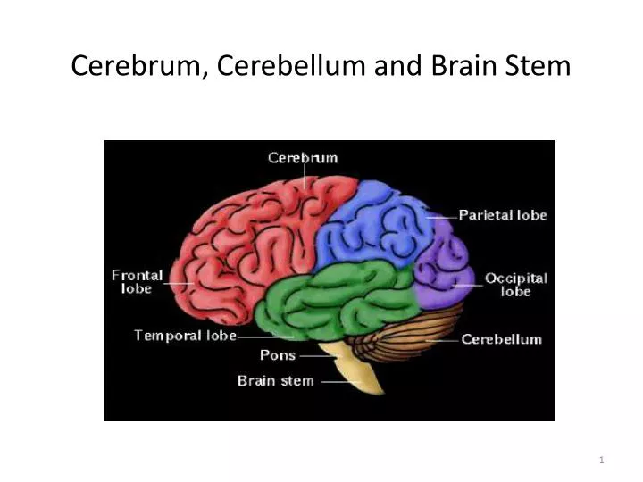
Cerebrum, Cerebellum and Brain Stem. C71.0. C71.3. C71.1. C71.4. C71.6. C71.2. C71.7. Meninges. CNS Cells. Two cell types Neuron Conducts nerve impulses Cannot be replaced if destroyed Neuroglial cells Support, nourish, and protect the neurons

E N D
Cerebrum, Cerebellum and Brain Stem C71.0 C71.3 C71.1 C71.4 C71.6 C71.2 C71.7
CNS Cells • Two cell types • Neuron • Conducts nerve impulses • Cannot be replaced if destroyed • Neuroglial cells • Support, nourish, and protect the neurons • Include astrocytes, oligodendrocytes, ependymal cells and microglia
Case No. 1 Meningioma
43 years old female complained of headache and two attacks of seizures in the past 4 months . Brain MRI revealed a 3 cm. extra-axial mass in the parietal region. It was dural based with mild edema in surrounding brain tissue. What is your provisional diagnosis?
A rounded mass attached to the dura compress the underlying brain but is easily separated from it. Meningioma arising from the dura at the base of the skull
A,parasagittalmultilobularmeningiomaattached to the dura with compression of underlying brain. B, Meningioma with a whorled pattern of cell growth and psammoma bodies.
MENINGIOMA(DURA-BRAIN) Meningioma:Section of tumour shows: • Whorls of fibrocellular tissue. • Cells are oval, spindle shape or elongated and lack mitosis. • Psammoma bodies (spherical calcified particles) are also seen within the tumour.
Case No. 2 Glioblastomamultiforme
55 years old man complained of headache for the last 2 months . Brain MRI reveals a 3 cm. frontal intra-parenchymalspace occupying lesion with rim enhancement on contrast studies. What is your provisional diagnosis ?
Computed tomographic(CT) scan of a large tumor in the cerebral hemisphere showing signal enhancement with contrast material and pronounced peritumoral edema. Glioblastoma multiforme appearing as a necrotic, hemorrhagic, infiltrating mass.
Glioblastoma. Foci of necrosis with pseudopalisading of malignant nuclei and endothelial cell proliferation Glioblastoma. Foci of necrosis with pseudopalisading of malignant nuclei.
Case No. 3 Multiple sclerosis
27 years old woman presents with a sudden onset of right sided blindness and weakness in her left leg. There is no history of trauma. However, she experienced a similar episode 8 months ago and was diagnosed as aseptic meningitis.
What is your provisional diagnosis? Multiple sclerosis. Section of fresh brain showing brown plaque around occipital horn of the lateral ventricle.
Multiple sclerosis. A, Unstained regions of demyelination (MS plaques) around the fourth ventricle (Luxol fast blue PAS stain for myelin). B, Myelin-stained section shows the sharp edge of a demyelinated plaque and perivascular lymphocytic cuffs. C, The same lesion stained for axons shows relative preservation.
This is a myelin stain (luxol fast blue/PAS) of an early lesion of pale demyelinated area. The lesion is centered around a small vein (arrow) which is surrounded by inflammatory cells . This is an H&E stained sections from a patient with long-standing MS. This lesion is centered on a vein. In this older lesion, however, there is very little inflammation around the vein. You can see the loss of myelin even without a special stain: it is lighter pink than the normal white matter surrounding it.
The key microscopic features of multiple sclerosis are: Perivenous mononuclear inflammation (lymphocytes, plasma cells and macrophages) Loss of myelin and variable loss of oligodendrocytes. Relative preservation of axons. Reactive astrogliosis (sclerosis). However, MS lesions do vary depending on their age. Early (acute) lesions are characterized by perivascular and parenchymal infiltration by inflammatory mononuclear cells, and myelin breakdown and phagocytosis by macrophages. Astrogliosis is not yet profound and axons are relatively preserved. As the lesion progresses, there are fewer inflammatory cells and more astrogliosis. Chronic lesions have few mononuclear cells, almost complete demyelination, and severe astrogliosis. There can be oligodendrocyte loss and some secondary axonal loss in advanced cases.
Case No.4 Schwannoma
39 years old man complains that he had noticed a progressive hearing loss over a 2 years period. Except for occasional headache, he has no other complaints . Evaluation discloses severe sensorineural hearing loss of the left side . MRI shows 1.5 cm. mass at the left cerebellopontineangle . What is your provisional diagnosis ?
Well circumscribed mass at the cerebello-pontine angle Schwannoma. A, Bilateral eighth nerve schwannomas. What syndrome is suggested by such finding?
A schwannoma. typically has dense areas called Antoni A (black arrow) and looser areas called Antoni B (blue arrows). The cells are elongated (spindle shaped) and the nuclei have a tendency to line up as seen here in the Antoni A area. A schwannoma is a benign tumor which can be cured by local excision . Schwannoma. B, Tumor showing cellular areas, including Verocay bodies (far right), as well as looser, myxoid regions.
Case No. 5 Hydrocephalus
9 months infant was suffering from enlarged head size and admitted to hospital with convulsions, went into coma and died. Autopsy was done and the brain was large with dilated ventricles . What is your provisional diagnosis?
Midsagittal magnetic resonance image of a child with communicating hydrocephalus, involving all ventricles. Hydrocephalus. Dilated lateral ventricles seen in a coronal section through the midthalamus. Cross section of brain showing marked dilatation of the ventricles.
Case No. 6 Pyogenic ( bacterial ) meningitis
4 years old child who was treated from otitis media and suddenly complained from headache, vomiting, fever and stiff neck. CSF was found to be clouded with abnormal increase of neutrophils, increased protein and absence of sugar. Gram stain of the CSF fluid showed meningiococci . What is your diagnosis ?
A thick layer of supurative exudate covers the brain hemispheres and meninges A thick layer of supurative exudate covers the brain hemispheres, with thickens Leptomeninges
Case No. 7 Brain abscess
35 years old lady complains from otitis media . Suddenly she suffers from headache and convulsions. Brain MRI reveals 5 cm. fluid filled cavity in the temporal lobe. Examination of the CSF shows increased pressure with lymphocytes and increased protein but there is no change of sugar content. What is your diagnosis ?
Multiple discrete lesions with central liquefactive necrosis. There is a surrounding fibrous capsule and marked edema
Case No. 8 Ruptured berry aneurysmcausing subarachnoid hemorrhage
A previously healthy 31-year-old woman experiences a severe headache and loses consciousness within an hour. An emergent head CT scan reveals extensive subarachnoid hemorrhage at the base of the brain. She is a febrile. A lumbar puncture yields cerebrospinal fluid with many red blood cells, but no white blood cells. The CSF protein is slightly increased, but the glucose is normal. What is your provisional diagnosis ?
View of the base of the brain, dissected to show the circle of Willis with an aneurysm of the anterior cerebral artery (arrow). Extensive hemorrhage in the pons with extension to fill the fourth ventiricle
B, Dissected circle of Willis to show large aneurysm. Common sites of saccular (berry) aneurysms in the circle of Willis C, Section through a saccular aneurysm showing the hyalinized fibrous vessel wall.
Case No.9 Alzheimer Disease
An 85 years old man complains of progressive loss of memory, disorientation and alterations in mood and behavior since 20 years. He was admitted to hospital because he was disabled and immobile and he died in hospital after one week of admission. Autopsy was done and the brain cortex was found to be atrophied. What is your diagnosis ?
Alzheimer disease with cortical atrophy most evident on the right, where meninges have been removed.
Alzheimer disease. A, Neuritic plaque with a rim of dystrophic neurites surrounding an amyloid core. Alzheimer Disease. C, Neurofibrillary tangles (arrowheads) are present within the neurons. Alzheimer Disease. D, Silver stain showing a neurofibrillary tangle within the neuronal cytoplasm.
Alzheimer Disease. B, Congo red stain of the cerebral cortex showing amyloid deposition in the blood vessels and the amyloid core of the neuritic plaque (arrow)
Alzheimer Disease • Macroscopic examination of the brain shows a variable degree of cortical atrophy with widening of the cerebral sulci that is most pronounced in the frontal, temporal, and parietal lobes • Microscopic: neuritic (senile) plaques, neurofibrillary tangles, and amyloidangiopathy