Download
1 / 41
410 likes | 431 Vues
PATHOLOGY OF THE UPPER RESPIRATORY TRACT Prof. Dr. Ferda ÖZKAN. Objectives. To learn the malformations of upper respiratory tractus To learn the inflammatory diseases of the upper respiratory tractus To learn the benign tumors of the upper respiratory tractus

E N D
PATHOLOGY OF THE UPPER RESPIRATORY TRACT Prof. Dr. Ferda ÖZKAN
Objectives • To learn the malformations of upper respiratory tractus • To learn the inflammatory diseases of the upper respiratory tractus • To learn the benign tumors of the upper respiratory tractus • To learn the malignant tumors of the upper respiratory tractus
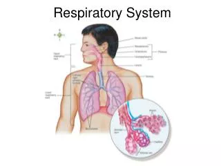
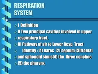
UPPER RESPIRATORY TRACT The term upper airways is usedto include the nose, pharynx, and larynx and their related parts.
CONGENITAL ANOMALIES • Cleft palate • Tracheo-esophageal fistulas • The newborn chokes and turns blue while eating. • In the severe forms, the esophagus begins in a blind pouch, and all food ends up in the airways.
NOSE & PARANASAL SINUSES • Inflammatory diseases, ‘common cold’ are the most common disorders of the nose and accessory air sinuses. • Most of the inflammatory diseases are of viral origin, but often complicated by superimposed bacterial infections.
Nose & Paranasal sinusesINFLAMMATIONS Infectious rhinitis“common cold” • caused by one or more viruses • adenoviruses, • echoviruses, • rhinoviruses. • They evoke a profuse catarrhal discharge.
These changes may extend, producing a concomitant pharyngotonsillitis. • Secondary bacterial infection mucopurulent or sometimes frankly suppurative exudate. • These infections soon clear up, usually in a week if appropriately treated but only after 7 days if ignored.
ALLERGIC RHINITIS (hay fever) • Initiated by sensitivity reactions to one of a large group of allergens, • most commonly the plant pollens, fungi, animal allergens and dust mites. • As is the case with asthma, allergic rhinitis is an IgE-mediated immune reaction. • The allergic reaction is characterized by marked mucosal edema, redness, and mucous secretion, accompanied by a leukocytic infiltration in which eosinophils are prominent.
NASAL POLYPS • Recurrent attacks of nonspecific or allergic rhinitis eventually lead to focal protrusions of the mucosa, • producing so-called nasal polyps, which may reach 3 to 4 cm in length.
Histologically, these polyps are not neoplasms, instead they consist of; • edematous mucosa having a loose stroma, • harbouring hyperplastic or cystic mucous glands, • inflammatory cells (prominently neutrophils, eosinophils, and plasma cells with occasional clusters of lymphocytes).
In the absence of bacterial infection, the mucosal covering is intact, but with chronicity, it may become ulcerated. • When multiple or large, they may enroach on the airway and impair sinus drainage.
CHRONIC RHINITIS • It is a sequel to repeated attacks of acute rhinitis either microbial or allergic origin with superimposed bacterial infection. • Histology reveals; - superficial desquamation or ulceration of the mucosal epithelium , - a variable inflammatory infiltrate of neutrophils, lymphocytes and plasma cells. • The suppurative infections sometimes extends into the air sinuses.
SINUSITIS • Acute sinusitis is most commonly preceded by acute or chronic rhinitis. • Maxillary sinusitis arises by extension of a periapical infection through the bony floor of the sinus. • Accused agents are usually inhabitants of the oral cavity. • Pooling of secretions enhance bacterial infection, contributing to accumulate the suppurative exudate, producing empyema of the sinus. • Edema (allergy, infection) around the outlets of the sinuses sets up a vicious cycle, with non-drainage followed by bacterial infection followed by additional obstruction.
Acute sinusitis may, in time, give rise to chronic sinusitis, particularly if there is interference with drainage. Usually there is a mixed microbial flora, largely of normal inhabitants of the oral cavity. Particularly severe forms of chronic sinusitis are caused by fungi (e.g., mucormycosis), especially in diabetics. Very uncommonly, sinusitis is a component of Kartagener’s syndrome, which also includes bronchiectasis and situs inversus. –All these features are secondary to defective ciliary action.
Necrotizing Lesions of the Nose and Upper Airways Necrotizing ulcerating lesions of the nose and upper respiratory tract may be produced by: • (1) spreading mucormycotic infections, particularly in the diabetics; • (2) Wegener’s granulomatosis • (3) lethal midline granuloma (polymorphic reticulosis).-in most cases a neoplasm of natural killer cells.
Nose & Paranasal sinusesTumors Sinonasal Papillomas • Benign neoplasms arising from the sinonasal mucosa and composed of squamous or columnar epithelium • HPV type 6 and 11 identified • Occur in three forms: -septal-(most common) -inverted (most important biologically) -cylindrical
Inverted papilloma • The inverted papilloma is a benign but locally aggressive neoplasm the papillomatous proliferation of squamous epithelium, • instead of producing an exophytic growth, it extends into the mucosa, so is called inverted • if not adequately excised, it has a high rate of recurrence, with the potentially serious complication of invasion of the orbit or cranial vault • rarely, frank carcinoma may also develop.
Isolated Plasmocytomas • These extramedullary plasmocytomas arise in the lymphoid structures adjacent to the nose and sinuses. • Polypoid growths varying from 1cm to several cms. covered by an intact overlying mucosa. • Histology is a malignant plasma cell tumor
Olfactory Neuroblastomas (Esthesioneuroblastomas) • Highly malignant tumors composed of small round cells resembling neuroblasts proliferating into lobular nests encircled by vascularized connective tissue. • They arise most often superiorly and laterally in the nose from the neuroendocrine cells dispersed in the olfactory mucosa.
Olfactory Neuroblastomas • Olfactory neuroblastomas tend to metastasize widely . • Combinations of surgery, radiation, and chemotherapy yield a 5-year survival rate of 50% to 70%.
Although the nasopharyngeal mucosa, related lymphoid structures, and glands may be involved in a wide variety of specific infections : diphtheria, infectious mononucleosis. NASOPHARYNX
Nasopharyngeal infections • Pharyngitis and tonsillitis are frequent concomitants of the usual viral upper respiratory infections: • rhinoviruses, • Echoviruses, adenoviruses, • respiratory syncytial viruses, • influenzal strains. • Bacterial infections may be superimposed on these viral involvements, or the bacteria may be primary invaders: • b-hemolytic streptococci, • Staphylococcus aureus or • other pathogens.
Particularly severe forms of pharyngitis and tonsillitis are seen • in infants and children who have not yet developed any protective immunity • neutropenia, • immunodeficiency, • uncontrolled diabetes, • disruption of the normal oral flora by antibiotics.
The inflamed nasopharyngeal mucosa may be covered by an exudative membrane (pseudomembrane), • the nasopalatine and palatine tonsils may be enlarged and covered by exudate • a typical appearance is of enlarged, reddened tonsils (owing to reactive lymphoid hyperplasia) • dotted by pinpoints of exudate emanating from the tonsillar crypts, so-called follicular tonsillitis.
The major importance of streptococcal “sore throats” lies in the possible development of poststreptococcal complications: • Rheumatic fever • Glomerulonephritis
Angiofibroma of the nasopharynx is usually a teenaged boy's tumor • it may cause serious clinical problems because of its tendency to bleed profusely during surgery.
Nasopharyngeal Carcinoma • Usually poorly-differentiated squamous cell carcinoma,seems to be caused by a combination of • (1) heredity, • (2) age, • (3) infection with EBV.
Nasopharyngeal carcinoma • Nasopharyngeal carcinomas take one of three patterns: • (1) keratinizing squamous cell carcinomas (WHO-1), • (2) nonkeratinizing squamous cell carcinomas (WHO-2), and • (3) undifferentiated carcinomas that have an abundant non-neoplastic, lymphocytic infiltrate (WHO-3). • The last pattern has often been called, erroneously, lymphoepithelioma.
LARYNX • Infections • The common cold and viral laryngitis. • Epiglottitis ("croup") usually results from infection with Hemophilusinfluenza B. (HIB) • Airway obstruction is the major problem. • Laryngeal papillomatosis is a life-threatening infestation of the larynx with HPV (strain 1).
Traumamost often results today from intubation. • Forensic pathologists look for fractures of the hyoid bone in suspected strangling. • Foreign bodies in the trachea can cause sudden death (in toddlers and drunken adults). • The "cafe coronary" is usually due to poorly-chewed beef. • Laryngeal nodules are benign fibroepithelial polyps on the vocal cords of those who use their voices a lot (singers, teachers; singers’ nodule). By convention, singers' nodules are bilateral lesions and polyps are unilateral. • They present as hoarseness and are easy to remove. • Laryngeal papillomas(solitary papillomas), usually occurring in adults, are true (benign) neoplasms, usually on the true vocal cords, that form soft, raspberry-like excrescences rarely more than 1 cm in diameter
Dysplasia of the laryngeal epithelium: • Pre-cancerousmore or less aggressive. • The range of squamous epithelial changes encountered in leukoplakia, erythroplakia, and squamous cell carcinoma of the oral cavity are replicated in the larynx. • Tuberculosis:looks like a laryngeal cancer in clinical examination. • Cancer of the larynx is very common • is almost invariably a squamous cell carcinoma due to cigarette smoking, • Ethanol abuse is also a risk factor, • Cancers confined to the true vocal cords (or the region just below them) are usually easy to cure using radiation, saving the voice, • Cancers above the true vocal cords usually require laryngectomy.
Comparison of a benign papilloma and an exophytic carcinoma of the larynx to highlight their quite different appearances. Layngeal lesions
Large, ulcerated, fungating lesion involving the vocal cord and piriform sinus. Laryngeal carcinoma
Histologic appearance of laryngeal squamous cell carcinoma. the atypical lining epithelium and invasive keratinizing cancer cells in the submucosa. Laryngeal carcinoma
Trachea Malignant tumors of the trachea • Rare • adenoid cystic carcinoma or • mucoepidermoid carcinoma • arising from tracheal glands that are homologous to salivary glands.