Download
1 / 66
680 likes | 857 Vues
Chapter 13 Nuclear Magnetic Resonance Spectroscopy. Leroy Wade. Origin of NMR Signals. Information contained in an NMR spectrum includes:. 1-Chlorobutane CH 3 CH 2 CH 2 CH 2 Cl. 1. Position (chemical shift) of the peaks – amount of shielding.

E N D
Chapter 13 Nuclear Magnetic Resonance Spectroscopy Leroy Wade
Information contained in an NMR spectrum includes: 1-Chlorobutane CH3CH2CH2CH2Cl 1. Position (chemical shift) of the peaks – amount of shielding. 2. Intensities of signals – number of protons producing each signal.
3. Splitting pattern – number of neighboring protons. • Number of signals – different types of proton present in the molecule. - protons that have different chemical shifts are chemically nonequivalent – exist in different molecular environment.
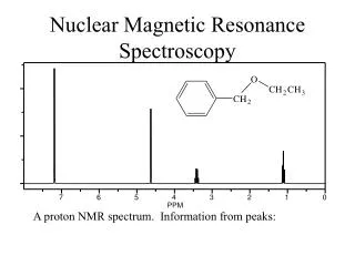
N CCH2OCH3 10.0 9.0 8.0 7.0 6.0 5.0 4.0 3.0 2.0 1.0 0 OCH3 NCCH2O Chemical shift (, ppm)
Chemically equivalent protons H3CCH2CH3 are in identical environments - have same chemical shift Replacement test: replacement by some arbitrary "test group" generates same compound chemically equivalent
Chemically equivalent protons ClCH2CH2CH3 CH3CH2CH2Cl Replacing protons at C-1 and C-3 gives same compound (1-chloropropane) C-1 and C-3 protons are chemically equivalent and have the same chemical shift H3CCH2CH3 chemically equivalent
2. CHEMICAL SHIFTS All protons present in a molecule don’t produce a single NMR signal. Protons in different chemical environments produce signal at different positions on the spectrum. For example, CH3CH2CH2CH2Cl produce a set of four signals, one for methyl and three for methylene protons. The position of signal’s appearance on NMR spectrum is known as Chemical Shift.
Measurement of Chemical Shift Position of resonance signals are measured relative to (CH3)4Si, tetramethylsilane (TMS), used as a NMR reference substance or standard. All 12 protons of TMS produce a single sharp line on NMR spectrum.
Resonance frequency of nuclei depends on the applied magnetic resonance frequency. In order to make the chemical shift values independent of the magnet strength, a ppm scale is introduced. This dimensionless chemical shift is represented by d is defined as follows.
Spin-Spin Splitting Not all NMR peaks are singlets. When two different types of protons are close enough their magnetic fields interact with each other and signals are splitted.
10.0 9.0 8.0 7.0 6.0 5.0 4.0 3.0 2.0 1.0 0 Cl2CHCH3 4 lines; quartet 2 lines; doublet CH3 CH Chemical shift (, ppm)
10.0 9.0 8.0 7.0 6.0 5.0 4.0 3.0 2.0 1.0 0 Cl2CHCH3 4 lines; quartet 2 lines; doublet coupled protons are vicinal (three-bond coupling) CH splits CH3 into a doublet CH3 splits CH into a quartet CH3 CH Chemical shift (, ppm)
H Cl H H C C H Cl Why do the methyl protons of1,1-dichloroethane appear as a doublet? To explain the splitting of the protons at C-2, we first focus on the two possible spin orientations of the proton at C-1 signal for methyl protons is split into a doublet
H Cl H H C C H Cl Why do the methyl protons of1,1-dichloroethane appear as a doublet? There are two orientations of the nuclear spin for the proton at C-1. One orientation shields the protons at C-2; the other deshields the C-2 protons. signal for methyl protons is split into a doublet
H Cl H H C C H Cl Why do the methyl protons of1,1-dichloroethane appear as a doublet? The protons at C-2 "feel" the effect of both the applied magnetic field and the local field resulting from the spin of the C-1 proton. signal for methyl protons is split into a doublet
H Cl H H C C H Cl this line correspondsto molecules in which the nuclear spin of the proton at C-1 reinforcesthe applied field this line correspondsto molecules in which the nuclear spin of the proton at C-1 opposesthe applied field "true" chemicalshift of methylprotons (no coupling)
H Cl H H C C H Cl Why does the methine proton of1,1-dichloroethane appear as a quartet? Theprotonat C-1 "feels" the effect of the applied magnetic field and the local fields resulting from the spin states of the three methyl protons. The possible combinations are shown on the next slide. signal for methine proton is split into a quartet
H Cl H H C C H Cl Why does the methine proton of1,1-dichloroethane appear as a quartet? There are eight combinations of nuclear spins for the three methyl protons. These 8 combinations split the signal into a 1:3:3:1 quartet.
The N+1 rule For simple cases, the multiplicity of a signalfor a particular proton is equal to the number of equivalent vicinal protons + 1.
Number of equivalent Appearance Intensities of linesprotons to which H of multiplet in multipletis coupled 1 Doublet 1:1 2 Triplet 1:2:1 3 Quartet 1:3:3:1 4 Pentet 1:4:6:4:1 5 Sextet 1:5:10:10:5:1 6 Septet 1:6:15:20:15:6:1 Splitting Patterns of Common Multiplets
Splitting Patterns:The Ethyl Group CH3CH2X is characterized by a triplet-quartet pattern (quartet at lower field than the triplet)
10.0 9.0 8.0 7.0 6.0 5.0 4.0 3.0 2.0 1.0 0 BrCH2CH3 4 lines; quartet 3 lines; triplet CH3 CH2 Chemical shift (, ppm)
Splitting Patterns:The Isopropyl Group (CH3)2CHX is characterized by a doublet-septet pattern (septet at lower field than the doublet)
10.0 9.0 8.0 7.0 6.0 5.0 4.0 3.0 2.0 1.0 0 7 lines; septet 2 lines; doublet CH3 CH Chemical shift (, ppm)
H H OCH3 Cl H H 10.0 9.0 8.0 7.0 6.0 5.0 4.0 3.0 2.0 1.0 0 skewed doublets OCH3 Chemical shift (, ppm)
Couplinmg Constants The distance between the peaks of a multiplet (in Hz) is called the Coupling Constant. Coupling constants are represented by J, and the coupling constant between Ha and Hb is represented by Jab.
Exercise: Fig 13-30
H H H O2N m-Nitrostyrene Consider the proton shown in red. It is unequally coupled to the protons shown in blue and yellow. Jcis = 12 Hz; Jtrans = 16 Hz
H H H O2N 16 Hz m-Nitrostyrene The signal for the proton shown in red appears as a doublet of doublets. 12 Hz 12 Hz
H H H O2N doublet doublet doublet of doublets
Time Dependence of NMR Spectroscopy Most conformational changes occur faster than NMR can detect them. An NMR spectrum is the weighted average of the conformations. For example: Cyclohexane gives a single peak for its H atoms in NMR. Half of the time a single proton is axial and half of the time it is equatorial. The observed chemical shift is half way between the axial chemical shift and the equatorial chemical shift.
1H NMR Spectra of O-H, N-H proton containing molecules The chemical shift for O—H and N-H is variable ( 0.5-5 ppm) and depends on temperature and concentration. Splitting of the O—H proton is sometimes observed, but often is not. It usually appears as a broad peak. Adding D2O converts O—H to O—D. The O—H peak disappears.
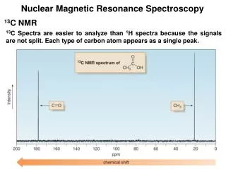
1H and 13C NMR compared: both give us information about the number of chemically nonequivalent nuclei (nonequivalent hydrogens or nonequivalent carbons) both give us information about the environment of the nuclei (hybridization state, attached atoms, etc.) it is convenient to use FT-NMR techniques for 1H; it is standard practice for 13C NMR
1H and 13C NMR compared: 13C requires FT-NMR because the signal for a carbon atom is 10-4 times weaker than the signal for a hydrogen atom a signal for a 13C nucleus is only about 1% as intense as that for 1H because of the magnetic properties of the nuclei, and at the "natural abundance" level only 1.1% of all the C atoms in a sample are 13C (most are 12C)